PLASMONICA 2026 Logo Plasmonica

Pisa, Italy — July 8–10, 2026

↶ Back to the main page ➜ Browse the program

List of Abstracts

Click an entry to expand the abstract.

Introducing the Degree of Collectivity in Lattice Resonances of Plasmonic Nanoparticle Arrays

V. Aglieri$^{(1)}$*, J. J. Alvarez-Serrano$^{(2)}$, M. Sohaib$^{(1,3)}$, J. R. Deop-Ruano$^{(2)}$, J. Olmos-Trigo$^{(4,5)}$, A. Toma$^{(1)}$, A. Manjavacas$^{(2)}$

  1. Istituto Italiano di Tecnologia, via Morego 30, 16163 Genova, Italy
  2. Instituto de Química Física Blas Cabrera (IQF), CSIC, 28006 Madrid, Spain
  3. Dipartimento di Fisica, Università degli Studi di Genova, via Dodecaneso 33, 16146 Genova, Italy
  4. Departamento de Óptica, Universidad Complutense de Madrid, 28040 Madrid, Spain
  5. Departamento de Física de Materiales, Universidad Autónoma de Madrid, 28049 Madrid, Spain

(*) vincenzo.aglieri@iit.it

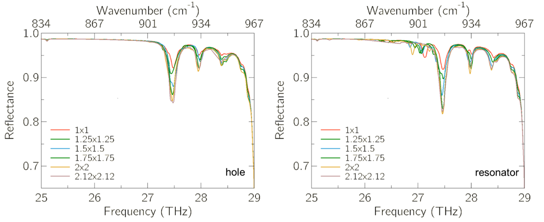
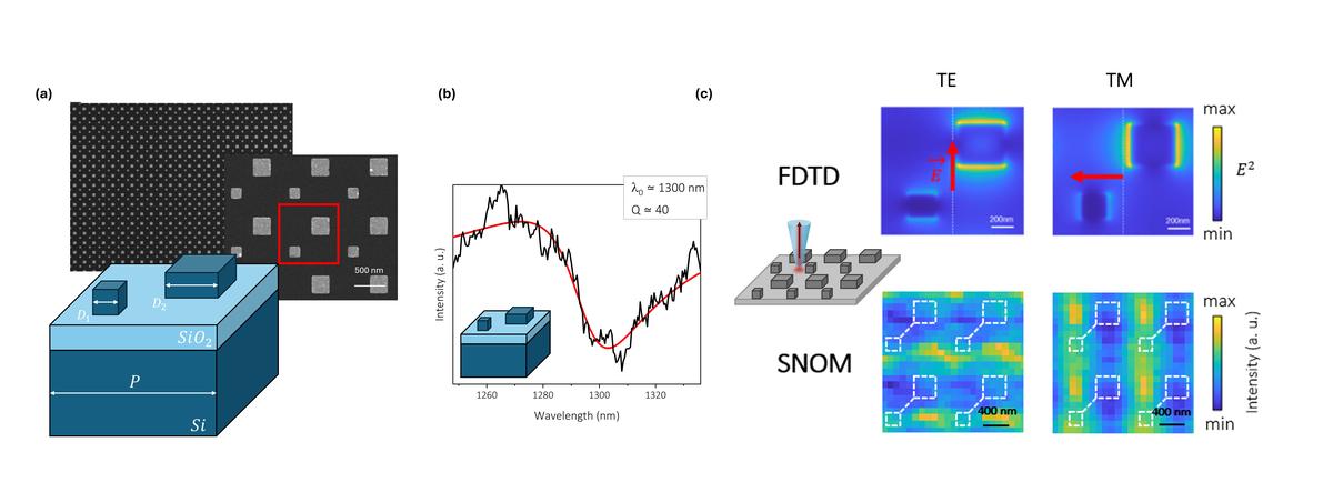
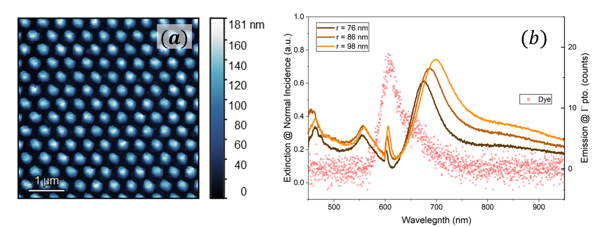
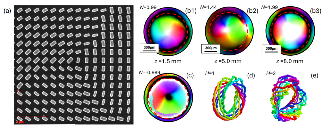
The excitation of localized surface plasmon resonances (LSPRs) on metal nanoparticles (NPs) has attracted much interest due to the possibility to concentrate light in nanometric volumes and reach high near-field enhancement. When NPs are arranged in array with periodicity comparable to the excitation wavelength, in-plane lattice resonance (LR) can emerge from the constructive interference of the individual NP response, showing extremely high Q-factor resonance with respect to the LSPR. As a result, LRs have found applications in many fields of study, including quantum information and photocatalysis [1]. Despite the increasing relevance of LR, a quantitative metric able to describe the strength of the interaction between NPs, and to guide the design of LR-supporting arrays, remains lacking. Here we present the recently introduced "degree of collectivity" [2], an experimentally accessible metric that quantifies the coherence of constructive interactions between NPs. By combining numerical and experimental studies, we demonstrate that higher degree of collectivity corresponds to higher LR quality-factor and NP array robustness against fabrication imperfection. For a more comprehensive understanding, we also evaluate the impact of the finite array size, periodicity imperfection, and environment refractive index on the LR behavior at a given degree of collectivity. Moreover, we experimentally demonstrate that the interplay between two NP arrays enables the excitation of out-of-plane modes at normal incidence [3]. Our results provide a reliable tool to predict the experimental performance and feasibility of actual plasmonic devices relying on LR, thus opening new scenarios in the realization of advanced photonic devices.

Support from the European Research Council under the European Union’s Horizon 2020 Research and Innovation Program through the ERC Consolidator Grant REPLY (Grant Agreement No. 101002422) is acknowledged.

References
  1. Kravets, V. G. et al. “Plasmonic surface lattice resonances: a review of properties and applications,” Chem. Rev., Vol. 118, No. 12, 5912–5951, 2018.3.
  2. Alvarez-Serrano, Juan J., et al. "Conceptualizing collectivity in lattice resonances of periodic arrays of nanostructures." JPhys Photonics 8.1 (2026): 015069
  3. Alvarez-Serrano, Juan J., et al. "Normal incidence excitation of out-of-plane lattice resonances in bipartite arrays of metallic nanostructures." ACS photonics 11.1 (2023): 301-309.

Nanophotonics for Biomedical Applications: From Chiral Optical Forces to Directional Photothermal Therapies

Guillermo Serrera$^{(1)}$, Javier González-Colsa$^{(1)}$, Alfredo Franco$^{(1)}$, José M. Saiz$^{(1)}$, Dolores Ortiz$^{(1)}$, Fernando Moreno$^{(1)}$, Francisco González$^{(1)}$, Fernando Bresme$^{(2)}$, Pablo Albella$^{(1)}$*

  1. Group of Optics, Department of Applied Physics, University of Cantabria, Spain.
  2. Department of Chemistry, Molecular Sciences Research Hub, Imperial College London, UK.

(*) pablo.albella@unican.es

Nanophotonics is opening new paradigms in biomedicine by enabling unprecedented control over light–matter interactions at the nanoscale. In this talk, we present recent advances in two complementary directions: chiral optical manipulation for enantiomer discrimination and thermoplasmonic strategies for targeted cancer therapy.

First, we address the challenge of enantioselective detection and separation, which is fundamentally limited by the weakness of chiroptical forces compared to dominant achiral interactions. We show how high-index dielectric nanostructures, specifically silicon nanodisks excited through structured light, can selectively enhance optical chirality gradients while suppressing unwanted field intensity variations. This approach enables robust enantioselective optical trapping, with predicted selectivities exceeding two orders of magnitude under realistic conditions, and extends the applicability of optical chirality to weakly chiral biomolecules.

In the second part, we explore nanoplasmonic platforms for photothermal therapy (PTT), where localized heating is used for selective cancer cell ablation. We introduce novel designs of nanoparticles, aimed at achieving both efficient light absorption and directional heat transport. By combining plasmonic heating with controlled thermal insulation, these structures generate strong temperature rises and directional heat flux, concentrating great part of the thermal energy toward a defined spatial region. This anisotropic heating mitigates off-target damage and enhances therapeutic precision.

Raman Spectroscopy with 1550 nm Excitation Enhanced by Optical Antennas

G. Ferrando$^{(1)}$, T. Venanzi$^{(2)}$, S. Sotgiu$^{(2)}$, G. Zambito$^{(1)}$, E. Fragomeni$^{(2)}$, M. Ortolani$^{(2)}$, F. Buatier de Mongeot$^{(1)}$, M.C. Giordano$^{(1)}$, L. Baldassarre$^{(2)}$*

  1. Dipartimento di Fisica, Università degli Studi di Genova, Via Dodecaneso 33, I-16146 Genova, Italy
  2. Dipartimento di Fisica, Sapienza University of Rome, P.le A. Moro 2, I-00185 Rome, Italy

(*) leonetta.baldassarre@uniroma1.it

The development of nanodevices working in the near-IR range is a key challenge for applications ranging from communications and energy conversion to sensing and diagnostics. In this context, two-dimensional (2D) transition metal dichalcogenide (TMD) semiconductors, with their strong light–matter interactions, provide an ideal platform for next-generation ultra-compact photonic technologies [1,2]. However, their intrinsically low optical absorption and limited quantum efficiency require advanced photonic strategies to enhance light coupling to ultrathin layers over. In order to develop hybrid 2D-antenna architectures, a thorough understanding of the possible losses due to scattering processes is essential. Raman spectroscopy with low-energy excitation is a powerful tool to probe the fundamental scattering properties of layered materials, although it typically suffers from extremely weak emission signals [3]. Here, we present 2D–plasmonic nanosystems designed to achieve resonant amplification of Raman emission with excitation in the infrared, enabling investigation of the optoelectronic response of 2D materials. We fabricate anisotropic plasmonic nanoantenna arrays with ultra-smooth surfaces using a thermal scanning probe nanolithography approach [4,5], demonstrating controlled excitation of localized plasmon resonances in the infrared. By conformally transferring few-layer MoTe$_2$ onto these nanoantennas, we realize hybrid 2D–TMD nanoarrays. Raman measurements performed at 1550 nm excitation, in resonance with the nanoantennas, show a significant enhancement of the otherwise weak infrared Raman signal, enabling measurements on few-layer MoTe$_2$. These results identify hybrid 2D–plasmonic nanosystems as a promising platform for enhancing weak photonic emissions in the infrared and pave the way for fundamental studies of 2D material properties.

References
  1. K.F. Mak and J. Shan, Nature Photonics, 4, 216 (2016).
  2. A. Varghese, Y. Yin, M. Wang, S. Lodha, N. V. Medhekar, Adv. Mater. Interfaces 9, 2102174 (2022).
  3. S. Sotgiu et al, Phys Rev B 113, 085201 (2026).
  4. M. C. Giordano et al. Adv. Mater. Interfaces 10, 5, 2201408 (2023).
  5. G. Ferrando et al. Small Science 5, 1, 2400272 (2025).

2D-ES Analysis of Colloidal Plexcitonic Nanohybrids

D. Baldon$^{(1)}$*, A. Inchingolo$^{(1)}$, F. Mancin$^{(1)}$, E. Collini$^{(1)}$

  1. University of Padova, Italy.

(*) elisabetta.collini@unipd.it

Polaritonic materials arise from strong coupling between the confined electric field of an optical cavity and the matter states of a quantum emitter (e.g., organic or inorganic excitons). They have attracted increasing interest for potential applications in photocatalysis, nanoscale energy transfer, and optoelectronics. Plexcitonic materials are a subclass in which plasmonic nanoparticles act as optical cavities through near-field confinement. These nanoparticles, synthesized in diverse morphologies via scalable wet-chemical methods, can be functionalized with selected emitters to couple to the enhanced plasmonic field. The coupling strength depends on plasmon–exciton detuning, dephasing rates, emitter–surface distance, and the number of coupled emitters. Regimes from weak to strong coupling can thus be accessed, exhibiting distinct dynamical behavior. Strong coupling occurs when coherent energy exchange between hybrid plexcitonic states exceeds dissipative losses. Understanding relaxation dynamics in this regime is essential for rational material design. Two-dimensional electronic spectroscopy (2D-ES) offers femtosecond resolution and excitation-frequency selectivity, making it a powerful tool for resolving ultrafast processes in strongly coupled systems. Here, plexcitonic systems were prepared using J-aggregates of the cyanine dye TDBC as quantum emitters and gold nanorods (AuNRs) as plasmonic nanocavities, whose longitudinal plasmon resonance was tuned to match the excitonic transition. The resulting strongly coupled systems were investigated by two-dimensional electronic spectroscopy (2D-ES) in the boxcars geometry, enabling separate acquisition of rephasing and non-rephasing spectra. 2D-ES measurements were performed on plexcitonic samples spanning different coupling regimes. For each sample, data were collected at varying pulse energies to probe potential power-dependent dynamical processes within the plexcitonic nanohybrids. In addition, the influence of the excitation laser spectrum was examined by conducting 2D-ES experiments at different excitation wavelengths, resonant with either the exciton reservoir or the lower plexciton branch. This selective excitation allowed different initial states to be preferentially populated by the first pulses, enabling us to investigate how the nature of the initially prepared state affects the subsequent ultrafast dynamics.

2D-ES Analysis of Colloidal Plexcitonic Nanohybrids
(a) TEM image of AuNRs and molecular structure of TDBC; (b) 2D-ES map of the AuNR/TDBC plexciton at waiting time 400 fs.

Enhancing Light–Matter Interactions with Plasmonic–Dielectric Nanostructures

A. Barreda$^{(1)}$*

  1. Group of Displays and Photonics Applications. Carlos III University of Madrid. Madrid 28911, Spain

(*) abarreda@ing.uc3m.es

Nanoscale antennas enable the confinement of electromagnetic fields into subwavelength regions, providing an effective route to boost the spontaneous emission of quantum emitters. In this context, a wide range of architectures based on metallic nanoparticles, high-index dielectrics, and their combinations have been investigated. In this work, we present a systematic comparison of planar metallic, dielectric, and hybrid nanoantennas, evaluating their performance in terms of three key metrics: Purcell enhancement, radiation efficiency, and emission directivity.

Our analysis focuses on dimer configurations composed of cylindrical nanoparticles with different material compositions and geometries. Among the studied cases, dimers formed by gold nanocylinders emerge as the most effective structures for enhancing spontaneous emission. In contrast to many previous studies that primarily address emission redirection within the plane of the nanostructure, we find that a configuration consisting of two relatively large gold cylinders predominantly radiates in the out-of-plane direction. This behavior is attributed to a strong electric quadrupolar contribution in the resonant response.

To further improve emission directionality, we incorporate silicon nanocylinders acting as directors, which leads to an increase in directivity by a factor of approximately 2.4 compared to the bare gold dimer. Based on these findings, we propose a hybrid platform combining gold and silicon nanoparticles, capable of both enhancing the emission rate of a single quantum dot and tailoring its radiation pattern.

The insights provided here may be relevant for applications in fluorescence enhancement and quantum photonics, particularly in the design of efficient single-photon sources based on quantum dots and other nanoscale emitters.

TiN nanohole arrays as integrated plasmonic NIR wavelength filter for multispectral imaging

Fritz Berkmann$^{(1)}$*, Sebastian Reiter$^{(1)}$, Jon Schlipf$^{(2)}$, Akant Sengül$^{(1)}$, Christian Mai$^{(2)}$, Christian Wenger$^{(2)}$, Inga Anita Fischer$^{(1)}$

  1. BTU Cottbus-Senftenberg Experimentalphysik und funktionale Materialien, Erich-Weinert-Straße, 03046 Cottbus, Germany,
  2. IHP - Leibniz Institut für innovative Mikroelektronik, Im Technologiepark 25, 15236 Frankfurt (Oder), Germany

(*) Fritz.Berkmann@b-tu.de

While polymer-based wavelength filters are standard for imaging applications in the VIS wavelength range, their performance at larger wavelengths is impacted by absorption losses. Alternatively, Faby Perot multilayer filters can be used, however, their fabrication is challenging. Here, plasmonic nanohole arrays can be a cost-effective alternative: in those structures, the spectral dependence of optical properties is tuned via lateral geometry adjustments, making their fabrication highly adaptable. Realising such filters in combination with Ge photodetectors on the Si platform provides a route towards cost-effective and scalable manufacturing. At the same time, the use of the Si platform imposes strict limitations on material choices. Here, we present current characterization results on Ge photodetectors in combination with TiN nanohole arrays for wavelength-selective photodetection. The devices were fabricated in a 200 mm Si technology line. While optical losses in TiN are larger compared to metals such as Ag or Au, leading to an increased spectral width of plasmonic resonances, deposition of the material with low surface roughness and structuring via standard etching processes is possible, enabling the highly scalable, reliable and reproducible fabrication of nanohole arrays. Such nanohole arrays support Fano shaped transmission peaks resulting from the interaction of surface plasmon polaritons and Rayleigh anomalies with a broad background. The spectral position of the transmission peak can be easily tuned via the array pitch. We discuss how an Ge photodetectors with different nanohole array geometries as filters and in conjunction with spectral reconstruction techniques can be used for multispectral imaging at NIR wavelengths on the Si platform.

Digital holography for the reconstruction of particles suspended in diamagnetic drops levitated in a magneto-gravitational trap

S. Biscotti$^{(1)}$*

  1. Independent research. Student at Sapienza University of Rome.

(*) biscottisofia4@gmail.com

The magneto-gravitational trap developed in this work enables the stable levitation of millimeter sized diamagnetic microdroplets, allowing for the non-invasive analysis of the particles suspended within them. Levitation is achieved by combining the gravitational potential with the magnetic contribution generated by anti-Helmholtz coils, which together create a local minimum of the total potential. The optical system is designed to provide uniform illumination of the levitating droplet: a beam modulated by an SLM passes through a half-wave plate, is split by a polarizing beam splitter, recombined with a pump beam, and expanded to illuminate the entire trapping region. A dichroic mirror directs the wavelength of interest toward the sample while transmitting the scattered light to the CCD, and a second laser provides the reference beam for digital holography. An ultrasonic horn, coupled with metal electrodes, introduces the droplet into the levitation zone and can generate controlled acoustic streaming to gently redistribute the internal particles before acquisition. The overall architecture integrates magnetic, acoustic, and optical manipulation of the droplet into a single system, allowing for precise control over both the sample position and the internal fluid conditions. This synergy enables the exploration of experimental configurations that would be impossible to achieve using conventional approaches. The three-dimensional reconstruction of the complex optical field is performed via Fresnel propagation in the paraxial regime, producing amplitude and phase maps at various propagation distances. To identify the plane where the suspended particles appear most sharply resolved, a focus metric based on the discrete wavelet transform is employed. This measurement exhibits a clear maximum at the focal plane, providing robust and computationally efficient axial localization. This conceptual framework lays the foundation for a new methodology dedicated to the three-dimensional characterization of particles and microstructures within levitating water droplets.

Digital holography for the reconstruction of particles suspended in diamagnetic drops levitated in a magneto-gravitational trap
Optical Layout of the Experimental Setup

Modelling exciton-polaritons within an extended Frenkel Hamiltonian framework

S. Botticelli$^{(1)}$*, E. Sangiogo Gil$^{(1)}$, G. Granucci$^{(1)}$

  1. Dipartimento di Chimica e Chimica Industriale, Università di Pisa, Pisa, Italia

(*) samuele.botticelli@phd.unipi.it

Exciton-polaritons are hybrid states arising from the strong coupling between molecular excitations and confined electromagnetic modes, such as those supported by plasmonic or Fabry-Pérot cavities [1]. The polaritonic regime can influence the properties of materials, promoting long-range energy and charge transfer [2] and modifying photorelaxation pathways, decay lifetimes, and quantum yields [3].

Here we present a surface hopping implementation based on an extended Frenkel Exciton Model for the simulation of exciton-polaritonic states and their nonadiabatic dynamics in multichromophoric systems. Built on the previous works of Sangiogo et al. [4], the model incorporates the photonic degrees of freedom and the light-matter coupling terms [5] with the exciton-polaritonic states expressed in a joint exciton-photonic basis.

To demonstrate the capabilities of this new framework, we applied it in the computation of the polariton dispersion spectra of an azobenzene aggregate under strong coupling conditions and in the simulation of the relaxation dynamics of the same aggregate after excitations in different spectral regions.

References
  1. R. Bhuyan et al., Chem Rev., 123, 10877-10919, (2023)
  2. D. J. Tibben et al., Chem Rev 123, 8044-8068, (2023)
  3. J. A. Hutchison et al., Angew. Chem. Int. Ed., 51, 1592-1596, (2012)
  4. E. Sangiogo et al., J. Chem. Theory Comput., 17, 7373-7383, (2021)
  5. J. Fregoni et al., J Comput Chem., 41, 2033-2044, (2020)

Lifshitz-like States in Hyperuniform Disordered Photonic Networks

G. Calusi$^{(1)}$*, N. Granchi$^{(1)}$, K. Stokkereit$^{(2)}$, M. Lodde$^{(3)}$, C. Gonzini$^{(1)}$, R.P.J. van Veldhoven$^{(3)}$, A. Fiore$^{(3)}$, M. Florescu$^{(4)}$, F. Intonti$^{(1)}$

  1. Department of Physics and Astronomy and LENS, University of Florence, Italy.
  2. Advanced Technology Institute and Department of Physics, University of Surrey, UK.
  3. Department of Applied Physics and Science Education, Eindhoven University of Technology, The Netherlands.
  4. Optoelectronics Research Centre, University of Southampton, UK.

(*) gabriele.calusi@unifi.it

Recently, Hyperuniform disordered (HuD) systems materials, intermediate between random media and ordered photonic crystals, have attracted growing interest as a versatile platform for light control. In these systems, disorder is no longer a limitation but a design resource. Here we investigate two-dimensional stealthy HuD dielectric networks (Fig.1a), generated via a tessellation protocol [1]. The degree of order, quantified by the stealthiness parameter $\chi$, ranges from fully random ($\chi = 0$) to nearly crystalline ($\chi \approx 1$). Despite their disordered nature, these systems support a photonic bandgap (PBG) [2]. Light transport is strongly frequency dependent: extended modes appear far from the PBG, while near the band edge modes become Anderson-localized over a few cells [2]. Additionally, defect-like states emerge within the PBG, strongly confined in four-sided cells (red in Fig.1a) due to topology. We demonstrate that these states are the photonic analogue of Lifshitz states in electronic systems [3]. Indeed, their number decreases with increasing $\chi$ (i.e. decreasing disorder), as shown in Fig.1b, a trend confirmed for different system sizes (N, number of cells). These modes exhibit strong spatial confinement and are spectrally near the band edge, consistent with the phenomenology of Lifshitz states, rare disorder-induced eigenmodes governing the density of states near band edges. However, unlike their electronic counterparts, photonic Lifshitz-like states are not rare statistical anomalies but predictable at the design stage, as they originate from four-fold defects of the network (Fig.1b inset). Finally, we show that if brought in closed spatial proximity, such states can couple. Finally, we show that when spatially close, these states can couple. Using Scanning Near-field Optical Microscopy (SNOM), we demonstrate for the first time in HuD dielectric networks the presence and coupling of Lifshitz-like states forming a “photonic molecule”. Through perturbative imaging, we reconstruct the local density of states, revealing large spectral splitting and spatial delocalization over two four-sided cells due to hybridization (Fig.1c). These results highlight a programmable disorder regime where localization, correlations, and coupling can be engineered, opening opportunities for random lasing, neuromorphic devices, and quantum photonics.

Lifshitz-like States in Hyperuniform Disordered Photonic Networks
Figure1: a) Sketch of the 24 x 24 $\mu$m$^2$ 2D theoretical design of the HuD network with N=4000 cells, and stealthiness $\chi$=0.5. The four-sided defects are highlighted in red. The cyan rectangle defines the region employed to decorate the experimental sample. b) Histogram of four-sided topological defects (sketched in the inset) in different realizations of HuD networks varying N (500, 1000, 10000 and 30000) and for increasing values of $\chi$, i.e. decreasing disorder. c) Experimental spectral shift maps of the coupled Lifshitz-like defects acquired in the black region of a).
References
  1. M. Florescu et al, PNAS 106, 49, (2009).
  2. N. Granchi et al, Adv. Opt. Mat., 10, 8, 2102565, (2022).
  3. N. Granchi, G. Calusi et al, accepted in Light Science & App.

Tunable superplanckian near-field radiative heat transfer in hyperbolic metamaterials

S. Campanaro$^{(1)}$*, L. Bursi$^{(1)}$, S. Curtarolo$^{(2)}$, A. Calzolari$^{(3)}$

  1. Università di Modena e Reggio Emilia, Italy
  2. Duke University, USA
  3. Istituto di Nanoscienze, CNR-NANO, Italy

(*) stefano.campanaro@unimore.it

Hyperbolic metamaterials (HMMs) have attracted significant attention due to their ability to support electromagnetic modes with arbitrarily large wavevectors, which are evanescent in conventional media. These high-k modes, known as volume plasmon polaritons, enable a dramatic enhancement of the photonic density of states, opening the way to phenomena such as sub-wavelength imaging, superlensing, and notably the superplanckian thermal radiation.

In this work [1], we present our latest results on near-field radiative heat transfer between hyperbolic metamaterial surfaces, demonstrating a controllable superplanckian energy exchange even in high temperature conditions. By engineering multilayer HMMs composed of alternating metallic and dielectric constituents, we exploit their extreme optical anisotropy: a metallic response along one axis and dielectric along the orthogonal direction to tailor the spectral and spatial properties of thermal emission.

A key aspect of our study is the introduction of tunability in the radiative heat flux, achieved through material design and external control parameters. This enables a transistor-like behaviour for thermal radiation, where the heat flux can be actively modulated as well as spectrally shaped. Such control goes beyond passive enhancement, providing a platform for dynamic thermal management at the nanoscale and could be useful to enhance the efficiency of thermophotovoltaic cells.

References
  1. S. Campanaro, L. Bursi, S. Curtarolo, A. Calzolari. Tunable superplanckian near-field radiative heat transfer in hyperbolic metamaterials, in preparation.

Babinet principle, inverse design and non-local effects: investigating phonon-polariton microstructures in the infrared region

E. Cannavò$^{(1)}$*, D. Baiocco$^{(1)}$, O. K. Jackson$^{(2)}$, Gonzalo Álvarez Pérez$^{(3)}$, E. Bozdogan$^{(4)}$, S. De Liberato$^{(2,4)}$, and A. Tredicucci$^{(1)}$

  1. Dipartimento di Fisica “E. Fermi”, Università di Pisa, Largo Pontecorvo 3, Pisa, Italy
  2. School of Physics and Astronomy, University of Southampton, Southampton, SO17 1BJ, United Kingdom
  3. Istituto Italiano di Tecnologia, Center for Biomolecular Nanotechnologies, Via Barsanti 14, 73010 Arnesano, Italy
  4. Istituto di Fotonica e Nanotecnologie – Consiglio Nazionale delle Ricerche (CNR), Piazza Leonardo da Vinci 32, Milano, Italy

(*) emmanuelecannavo@gmail.com

The usage of Surface Phonon Polaritons (SPhPs) in nanophotonics has demonstrated great promise for sub-wavelength light confinement in polar dielectrics. The significant reduction of optical losses and wide availability of polar crystals makes SPhPs a viable alternative to plasmonic systems. These modes can be excited between the longitudinal optical and transverse optical phonon frequencies, in the so-called Reststrahlen band of the dielectric, which, for most materials of interest, spans the Terahertz or mid-infrared ranges of the spectrum. Here we present the electromagnetic theory of different classes of phonon polariton metamaterials and hybrid structures, highlighting peculiar aspects of their dielectric response. In hole/pillar photonic crystals we discuss the cross-over between 3D and quasi-2D systems, demonstrating that, in the latter case, a version of the Babinet principle emerges, connecting the optical properties of complementary structures. In the context of graphene/SrTiO3 interfaces we investigate the importance of non-locality of the 2D electronic response to properly model coupled plasmon-phonon polaritons. Phonon non-locality becomes instead relevant in nanometer size particles, deeply modifying the usual Mie scattering cross section. An effective frequency-dependent local dielectric constant approach is shown here to give reliable, quick predictions, and experiments are also proposed to reveal the presence of such non-local contributions. Finally, we showcase a deep learning (DL) model aimed at inverse-designing a phonon polariton metamaterial whose absorption spectrum is matched to the spectral signature of a specific chemical substance in its gaseous or liquid state. To this end, we employ a simple model of a SiC (Reststrahlen band from 25 to 29 THz) micro-array and apply the DL algorithm. Data generation is carried out by producing absorption spectra of SiC nanostructures, like the ones showed in the attached figure, varying the pillars’ dimensions and the parameters of the unit cell of the periodic array. The simulated spectra are computed using the Electromagnetic Waves, Frequency Domain module of the COMSOL Multiphysics package. For the training process, a direct, one-shot network is adopted, in the form of a modified conditional variational autoencoder. The loss function depends on the overlap between the absorption spectra of the SiC microstructure and of the chemical species of interest, in proximity of its dominant spectral signature.

We acknowledge financial support under the National Recovery and Resilience Plan (NRRP), Mission 4, Component 2, Investment 1.1, Call for tender No. 1409 published on 14.9.2022 by the Italian Ministry of University and Research (MUR), funded by the European Union – NextGenerationEU – Project: Mid-Infrared Nanophotonics for Sustainability (MINAS) – CUP I53D23006670001 - Grant Assignment Decree No. 1381 adopted on 1.9.2023 by the Italian Ministry of of University and Research (MUR).

Babinet principle, inverse design and non-local effects: investigating phonon-polariton microstructures in the infrared region
Simulated reflectance spectra for square-based, 0.1 $\mu$m-high/deep resonator (right)/hole (left) SiC periodic arrays for different base edge lengths. Square unit cell with 3 $\mu$m edge.

All-optical investigation of chirality-driven interactions of spherical gold nanoparticles

G. Capizzi$^{(1,2)}$*, B. Fantasia$^{(1)}$, A. Capocefalo$^{(1,3)}$, C. Fasolato$^{(1,3)}$

  1. Department of Physics, Sapienza University, Rome, Italy
  2. Department of Basic and Applied Sciences for Engineering, Sapienza University, Rome, Italy
  3. Institute for Complex Systems, CNR, Rome, Italy

(*) giovanna.capizzi@uniroma1.it

Noble metal plasmonic nanoparticles (NPs) exhibit a strong interaction with visible light due to their surface plasmon resonance, i.e. the collective oscillation of their free electrons, which can be radiatively excited. For this reason, in the last decades Ag and AuNPs have been widely exploited as high-performance colorimetric [1] and spectroscopic [2] optical sensors. These systems can be easily functionalized on-demand through thiol-chemistry, offering great interface tunability. The perspective exploitation of plasmonic NPs as chiral sensors is particularly appealing, due to their potential ultra-high sensitivity, necessary in many applications (e.g., enantiomeric discrimination in pharmacology). In this context, relevant efforts are devoted to synthesizing AuNPs with chiral shapes [3]. Here, we focus on commercially available spherical AuNPs synthesized via the well-known citrate reduction protocol. We investigate the interaction of the same AuNP batch with different chiral organic analytes by UV-Visible and surface-enhanced Raman scattering (SERS) spectroscopy. Despite the spherical (i.e., achiral) shape of AuNPs, we surprisingly observe a selective interaction with a specific enantiomer, which is detectable colorimetrically through NP aggregation, causing the appearance of red-shifted components in the localized surface plasmon resonance spectrum. Zhang et al [4] hypothesized that this enantioselective interaction originates from the structural properties of the NPs (crystallite arrangement within the core). We instead highlight the role of the AuNP molecular stabilizing interface as a source of asymmetry. By SERS measurements, we show that pro-chiral citrate molecules bind asymmetrically to the metal surface, acting in facts as a chiral interface. To further explore this aspect, we modify the interface by substituting the citrate layer with achiral and chiral stabilizers, as well as considering a different AuNP size. We find that the achiral molecular interface leads to a loss of the chiral selectivity (Figure 1), while the chiral ones lead to a programmable enantioselective interaction with the organic molecules. This observation is remarkable, as it rules out an intrinsic enantioselectivity of the AuNPs, and it rather demonstrates that enantioselectivity can be introduced in plasmonic NPs by post-synthesis functionalization methods. By probing interparticle interactions through optical spectroscopy and investigating NP surface chemistry via SERS, we establish a general description of chirality-driven interactions in plasmonic nanosystems.

All-optical investigation of chirality-driven interactions of spherical gold nanoparticles
Figure 1: Rationale of the experiment. Colloidal spherical AuNPs interact with chiral analytes (S and R enantiomers) via long-range electrostatic and short-range weak interactions mediated by the surface interface. (a) Prochiral citrate ions bind asymmetrically to the gold surface, creating an effective chiral system and inducing enantioselective aggregation with distinct red-shifted features. (b) An achiral interface suppresses this effect, yielding no selectivity. (c) A chiral interface enables programmable enantioselectivity.
References
  1. Aldewachi, et al. Nanoscale 10.1 (2018): 18-33.
  2. Fasolato, et al. Nanoscale 8.39 (2016): 17304-17313.
  3. Lee, et al. Nature 556.7701 (2018): 360-365.
  4. Zhang, et al. Analytical Chemistry 95.14 (2023): 6107-6114.

Re-entrant plasmon coupling in thermoresponsive colloids

A. Capocefalo$^{(1,2)}$, Francesco Brasili$^{(1,2)}$, Emanuela Zaccarelli$^{(1,2)}$, Domenico Truzzolillo$^{(3)}$, Simona Sennato$^{(1,2)}$

  1. Institute for Complex Systems, CNR, IT
  2. Physics Dept., Sapienza University of Rome, IT
  3. Laboratoire Charles Coulomb, CNRS-Université de Montpellier, FR

(*) capocefalo.a@gmail.com

The development of flexible photonic devices with tunable optical properties has attracted increasing attention in recent years. A well-established strategy to integrate photonic components into soft materials relies on including plasmonic nanoparticles (NPs) within thermoresponsive microgels. In these hybrid particles, the volume phase transition (VPT) of the polymeric network, leading to a substantial microgel shrinking, links microscale structural changes to near-field plasmon interactions, enabling external control over interparticle distances and plasmon coupling. However, the extent to which the optical response can be programmed in these systems remains underexplored, particularly with respect to colloidal stability, despite its potential to give rise to a much broader range of optical behaviors. We reveal a counter-intuitive re-entrant behavior in the optical properties of electrostatically assembled microgel–NP complexes depending on the NP-to-microgel number ratio. Increasing NP loading initially enhances plasmon coupling, with a progressive redshift and broadening of the optical response across the VPT, but beyond a critical threshold the optical response narrows again toward that of weakly interacting NPs. To rationalize this behavior, we combine optical spectroscopy with X-ray and dynamic light scattering techniques to establish a quantitative relationship between plasmonic coupling and interparticle distance, identifying two distinct coupling regimes. We then demonstrate that, beyond the VPT-driven gathering of NPs within individual complexes, plasmonic coupling is also governed by inter-complex interactions that emerge at intermediate NP loadings, where loss of colloidal stability leads to the formation of large aggregates and to pronounced enhancement of the plasmonic response. We rationalize the overall phenomenology within the framework of charge-patch interactions by constructing a NP-loading-temperature phase diagram that directly links colloidal stability to optical response. Our findings demonstrate that plasmonic coupling in soft colloidal systems is controlled not only by nanoscale structure but also by mesoscale phase behavior, thus establishing a general framework for harnessing colloidal interactions to program optical responses in soft photonic materials.

Liposome-Gold Nanoparticles hybrid for Reproducible SERS Tags in Bioanalytical Applications

J. Cardellini$^{(1)}$*, C. Dallari$^{(2,3)}$, C. Credi$^{(2,3)}$, F.S. Pavone$^{(2,3)}$, A. Morrone$^{(4)}$, M. Calamai$^{(2,3)}$, C. Montis$^{(1)}$, D. Berti$^{(1)}$

  1. Department of Chemistry “Ugo Schiff” and CSGI, University of Florence, Florence, Italy
  2. National Institute of Optics—National Research Council, Sesto Fiorentino, Italy
  3. European Laboratory for Non-Linear Spectroscopy (LENS), Sesto Fiorentino, Italy
  4. Department of Neurosciences, Psychology, Drug Research and Child Health (NEUROFARBA), University of Florence, Florence, Italy

(*) jacopo.cardellini@unifi.it

Gold nanoparticles (AuNPs) interacting with lipid vesicles are widely studied as model systems to investigate nano–bio interfacial phenomena [1]. Yet, the potential of lipid membranes to actively template AuNPs assembly for functional applications remains largely unexplored. In this framework, we recently discovered that liposome membranes can actively direct the clustering of AuNPs, yielding AuNP–liposome hybrids whose size, organization, and colloidal stability are dictated by lipid composition and concentration [2]. This strategy shifts the role of the membrane from a passive support to an active regulator of mesoscale organization. We exploit this concept to address a long-standing challenge in surface-enhanced Raman scattering (SERS) [3]. SERS application remains limited by the challenging preparation of SERS tags, i.e. metallic nanoparticles functionalized with Raman-active molecules (RRs). Due to the hydrophobic nature of RRs, conventional SERS tags generally suffer from low colloidal stability in water, poor reproducibility, and their fabrication involves complex synthetic procedures. We introduce LipoGold Tags, hybrid SERS tags formed by simple self-assembly of AuNPs on liposome membranes. In this architecture, the lipid bilayer simultaneously encapsulates RRs and induces AuNP clustering, positioning the RRs within plasmonic hot spots [4]. Compared to existing SERS tags, LipoGold Tags exhibit enhanced colloidal stability, high reproducibility, and modular functionalization with targeting ligands. As a proof of concept, we demonstrate their application in the sensitive detection of infantile GM1 gangliosidosis, outperforming standard fluorescence-based assays. Overall, this work advances the field by demonstrating how soft membrane templating can be leveraged to rationally design functional plasmonic nanomaterials for bioanalytical applications.

References
  1. Caselli, L., et al. Nanoscale Horizons (2025).
  2. Cardellini, J., et al. Journal of the American Chemical Society (2025)
  3. Schumacher, M., et al. Advanced Optical Materials (2022)
  4. Cardellini, J., et al., Nature Communications (2024)

Mid-IR SESAMs for fiber lasers mode-locking at 3.6 um based on intersubband transitions in strong coupling

S. Cassandra$^{(1)}$*, E. Cosentino$^{(1)}$, A. Mukherjee$^{(1)}$, R. Riccio$^{(1)}$, S. Pirotta$^{(1)}$, J.-M. Manceau$^{(1)}$, A. Bousseksou$^{(1)}$, M. Jeannin$^{(1)}$, R. Colombelli$^{(1)}$, M. Malerba$^{(2)}$, S. Hatim$^{(3)}$, K. Eremeev$^{(3)}$, A. Hideur$^{(3)}$, M. Fagot$^{(4)}$, L. Cerutti$^{(4)}$, J.-B. Rodriguez$^{(4)}$, T. Berthelot$^{(5)}$, S. Cozic$^{(5)}$, S. Poulain$^{(5)}$

  1. Centre for Nanosciences and Nanotechnologies, UMR 9001 CNRS and Université Paris-Saclay, 91120 Palaiseau, France
  2. Istituto Nazionale di Ricerca Metrologica (INRIM), Strada delle Cacce 91, 10135 Torino, Italy
  3. CORIA UMR6614, CNRS-INSA-Université de Rouen, Normandie Université, 76801 Saint Etienne du Rouvray, France
  4. Institut d’Électronique et des Systèmes, Université de Montpellier, CNRS, F-34000 Montpellier, France
  5. Le Verre Fluoré, rue Gabriel Voisin, Campus de Ker-Lann, 35170 Bruz, France

(*) simone.cassandra@cnrs.fr

Laser mode-locking is an established technique for generating sub-ps pulses. In particular, self-starting mode-locking typically relies on saturable absorbers (SA): periodical self-modulation of intra-cavity losses locks the phases of longitudinal lasing modes and ultra-short pulses emerge from constructive interference [1]. SESAMs (semiconductor SA mirrors) represented a major breakthrough: quantum wells (QW) within mirrors induce a non-linear reflectivity increase with laser intensity by bleaching an interband absorption, enabling sub-ps pulses in solid state and semiconductor lasers [1]. While effective for lasers in the visible/NIR, an extension to the mid-IR (3-30 $\mu$m) is lacking due to photon energies below typical semiconductor bandgaps. Here, we present a semiconductor-based platform that extends SESAMs in the mid-IR. We target mode-locking of fiber lasers emerging in the 1st atmospheric window (3-5 $\mu$m) [2]. Our devices leverage ultrafast switching of strong light-matter coupling between intersubband (ISB) transitions in QWs and metal-dielectric-metal resonators [3, 4]. In this system, light-matter coupling is engineered for operation in strong coupling regime. When an intense laser beam illuminates the system, the ISB transition saturates, driving the system into weak coupling, which is characterized by a higher reflectivity value. This translates into a SESAM-like non-linear reflectivity, where the ultrafast ISB lifetimes provide the ps-level dynamics required for SESAM operation [5]. The presented platform permits to engineer the main figures of merit, such as modulation depth, linear reflectivity and saturation fluence via quantum design of the heterostructure and electromagnetic design of the resonator array. Preliminary experiments have been conducted on a recently developed Er-doped ZBLAN fiber laser (gain region 3.3-3.7 $\mu$m, peak gain 3.47 $\mu$m) [2]. The laser emission was tuned across the SESAM modulation region via a diffraction grating mounted in Littrow configuration, with the SESAM as a cavity mirror. Self-starting pulses are observed across the modulation bandwidth (3.6-3.7 $\mu$m), confirmed by optical spectrum broadening ($\Delta \lambda$ from 0.4 nm to 1.2 nm) and an RF beatnote - with its harmonics - at frep =22 MHz. The system shows temporal stability, confirming robustness and absence of laser-induced damage. Future experiments will focus on 2nd order autocorrelation, to reveal whether this is true soliton mode-locking or Q-switch mode-locking. Additionally, we are implementing SESAMs centered on the laser's peak gain. This optimization will eliminate the need for the diffraction grating, potentially enabling further spectral broadening towards transform-limited ultrashort pulses.

Mid-IR SESAMs for fiber lasers mode-locking at 3.6 um based on intersubband transitions in strong coupling
a) SEM image of MIR-SESAM, with typical cavity configuration. b) Non-linear SESAM reflectivity. c) SESAM ultra-fast temporal dynamics. d) RF Spectrum in pulsed operation. e) Optical spectrum of CW and mode-locked regime.
References
  1. U. Keller, Ultrafast Lasers, Springer (2022)
  2. S. Hatim. et al., Journal of the European Optical Society (2025)
  3. M. Jeannin, et al., Physical Review Letter (2021)
  4. M. Jeannin, et al., Applied Physics Letters (2023)
  5. M. Knorr, et al., Physical Review Letters (2022)

Broadband light harvesting with flat-optics 2D TMD-plasmonic nanoarrays

R. Chennuboina$^{(1)}$*, G. Ferrando$^{(1)}$, G. Zambito$^{(1)}$, M. Barelli$^{(1)}$, M.C. Giordano$^{(1)}$, F. Buatier$^{(1)}$

  1. University of Genoa

(*) rajesh.chennuboina@edu.unige.it

Two-dimensional Transition Metal Dichalcogenides (2D-TMDs) have attracted considerable interest due to their exceptional, layer-dependent optoelectronic properties, strong visible-light interaction, and strain tunability. Although their absorption exceeds that of bulk semiconductors by orders of magni-tude, it declines to ~10% in the ultra-thin limit and becomes negligible in the near-infrared, limiting their nanophotonic applicability. In this work we develop large-area flat-optics nanostructure arrays based on hybrid 2D TMDs - plasmonic nanoantenna’s, demonstrating superior photon harvesting prop-erties over a broadband Visible and Near-Infrared spectrum. A novel nano-fabrication technique based on the light interference lithography in back-etch configuration is exploited to create flat-optics nanoar-rays based on few-layer MoS2 to engineer their optical response. These subwavelength nanoarrays strongly enhance the Visible optical absorption thanks to the excitation of Rayleigh anomalies that also promote light scattering via guided mode resonances. To further improve the light coupling to these flat-optics layers we engineered metal–semiconductor–metal (MSM) architectures based onto the MoS$_2$ nanoarrays, with subwavelength lateral resolution. Maskless deposition of periodic Au and Al nano stripe arrays decorating the few-layer MoS$_2$ nanostripes is achieved through glancing angle evapora-tion, thus fully preserving the TMD crystal integrity while enabling lateral disconnection of the metal-lic nanostripes. Under this configuration hybrids photonic–plasmonic modes are excited promoting light-matter interaction and guiding light parallel to the ultra-thin high refractive index TMDs layers. This led to an enhanced optical absorption, exceeding 50% of the MSM nanoarrays with respect to the bare TMDs nanostructures. Remarkably, this effect is observed over an ultra-broadband spectral range across the Visible and the Near-Infrared. Therefore, the plasmonic integration with excitonic function-alities in two-dimensional semiconductors, offers a new opportunity for light-harvesting and optoelec-tronic devices.

Modelling plasmon-mediated photocatalysis

Leonardo Biancorosso$^{(1)}$, Giulia Dall'Osto$^{(2)}$, Margherita Marsili$^{(3)}$, Mauro Stener$^{(1)}$, Stefano Corni$^{(4)}$, Emanuele Coccia$^{(1)}$*

  1. Dipartimento di Scienze Chimiche e Farmaceutiche, University of Trieste
  2. Elettra Sincrotrone Trieste
  3. Dipartimento di Fisica e Astronomia "Augusto Righi", University of Bologna
  4. Dipartimento di Scienze Chimiche, Universit`a di Padova & Istituto Nanoscienze-CNR, Italy

(*) ecoccia@units.it

The interaction of light with matter is the subject of boundless research, ranging from astrophysics to materials science to biology and medicine. Theory is essential to interpret experimental outcomes and predict new features from, e.g., photophysics, photochemistry and photocatalytic studies. Clearly, time-dependent quantum mechanics is the appropriate tool for that purpose, allowing one to model fast and ultrafast dynamics occurring in chemical systems, and to properly simulate spectroscopy. Among the countless fields of research in which the role of light plays a key role in inducing transformations, molecular nanoplasmonics has seen growing interest develop around it, both experimentally and theoretically. Molecular nanoplasmonics [1,2] exploits collective electron excitations in metal nanostructures to enhance and control properties of molecules under the influence of light. A theoretical multiscale time-domain approach [3] was applied to study the selectivity toward methane in the photocatalytic hydrogenation of carbon dioxide in the presence of a rhodium nanocube [4,5] and to understand the dependence of hydrogen production rate on the extinction spectrum of a gold nanorod in the dehydrogenation of formic acid on palladium [6,7]. In both cases, we simulate the ultrafast plasmonic effects on the photoinduced charge distribution of the molecular stable intermediates of the reaction, thus predicting the fate of the processes and explaining the experimental outcomes.

References
  1. L. Novotny and B. Hecht, Principles of Nano-Optics, Cambridge University Press (2012)
  2. A. Lauchner et al., Nano Lett., 15, 6208 (2015)
  3. E. Coccia, J. Fregoni, C. A. Guido, M. Marsili, S. Pipolo and S. Corni, J. Chem. Phys., 153, 200901 (2020)
  4. X. Zhang et al., Nat. Commun., 8, 14542 (2017)
  5. G. Dall’Osto, M. Marsili, M. Vanzan, D. Toffoli, M. Stener, S. Corni and E. Coccia, J. Am. Chem. Soc., 146, 2208 (2024)
  6. Z. Zheng, T. Tachikawa and T. Majima, J. Am. Chem. Soc, 137, 948 (2015)
  7. L. Biancorosso and E. Coccia, J. Phys. Chem. Lett., 16, 12931 (2025)

Vibrationally Mediated Coherent Energy Exchange in Plexcitonic Nanomaterials

Federico Toffoletti$^{(1)}$, Giovanni Parolin$^{(2)}$, Dario Baldon$^{(1)}$, Giulia Dall’Osto$^{(3)}$, Lucio Litti$^{(1)}$, Stefano Corni$^{(1,4)}$, Elisabetta Collini$^{(1)}$*

  1. Department of Chemical Sciences, University of Padova
  2. Interdisciplinary Center for Scientific Computing, Ruprecht-Karls University
  3. Elettra Sincrotrone Trieste
  4. CNR-NANO Institute of Nanoscience

(*) elisabetta.collini@unipd.it

Harnessing strong light–matter coupling to manipulate molecular dynamics is a central goal in modern photonics. Hybrid light–matter states—plexcitons—arising from the interaction between plasmonic and excitonic modes provide a powerful route toward this goal. However, how molecular vibrations influence their coherent dynamics remains elusive. Here, we use femtosecond two-dimensional electronic spectroscopy to directly resolve vibronic interactions in plexcitonic nanostructures formed by cyanine molecular aggregates coupled to gold nanorods. We observe long-lived quantum beatings that evolve from vibronic plexcitonic coherences into vibrational coherences of predominantly excitonic character following plasmonic relaxation. This behavior reveals that molecular vibrational modes are not quenched under strong coupling but instead actively sustain coherent energy exchange across the disordered plexcitonic manifold, particularly between the lower plexciton and the exciton reservoir region. The excitonic component supports vibronic coherence formation, while the plasmonic admixture facilitates their generation and delocalization. Our findings identify vibronic coupling as a central mechanism governing light–matter interactions at the nanoscale, unveiling new routes to control coherence, Raman activity, and energy transport in strongly coupled plexcitonic nanostructures.

Sputtering-Based Solid-State Dewetting for integrating plasmonic structures on the facet of optical fibers

A.Corrado$^{(1,2)}$*, L.Piscopo$^{(1,2)}$, J.Pacella$^{(1,2)}$, S.Milanese$^{(1)}$, L.Fachechi$^{(1)}$, F.Rizzi$^{(1)}$, F.Pisanello$^{(1,\dagger)}$, M.De Vittorio$^{(1,2,3,\dagger)}$

  1. Istituto Italiano di Tecnologia, Center for Biomolecular Nanotechnologies, Arnesano, LE 73010, Italy
  2. Dipartimento di Ingegneria dell’Innovazione, Università del Salento, Lecce 73100, Italy
  3. Department of Health Technology, Drug Delivery and Sensing Section (IDUN), Technical University of Denmark (DTU), Kogens Lyngby 2800, Denmark

(*) alessandra.corrado@iit.it

The integration of plasmonic nanostructures onto optical fiber facets represents a transformative approach for integrating high efficiency optical biosensing into very small systems, accessing applications that include surface-enhanced Raman scattering (SERS) [1], refractive index (RI) sensing [2], surface-enhanced infrared absorption (SEIRA) [3], plasmon-enhanced fluorescence (PEF) [4]. Despite their potential, the widespread adoption of these on-fiber technologies is hindered by the low throughput of conventional nanofabrication methods. For instance, focused ion beam (FIB) milling [5-6] can be employed to pattern gold-coated fibers and fabricate periodic arrays of nanoplatles and nanograting, with the drawback of ion doping contamination of the facet. E-beam lithography overcome this disadvantage, but processing large-area nanostructures remains still a challenge [6-7]. Despite their high resolution, these methods require hours of processing to prepare a single plasmonic optical fiber and cannot be applied in parallel to multiple samples. To increase throughput, the scientific community has proposed a set of alternative methods, including electrostatic self-assembly directly in the fiber facet [8], nanoimprint lithography (NIL) [9], and solid state dewetting (SDW) starting from a thin metallic film. In particular, SDW requires the deposition of thin layers of gold (from few to tens of nanometers) followed by thermal processing at high temperatures ($\sim 600^\circ C$) to spontaneously nucleate metallic nanoislands (NIs). Despite the obtained random patterns, this method allows for plasmonic resonances for sensing applications and localized thermoplasmonic heating [10-11] with a process that can be simply run on tens of fibers simultaneously. Here we propose a study on SDW fabrication approach based on sputter-coating instead of electron-beam deposition. Unlike evaporation, sputtering enables a 3D clusters growth mode during deposition (Figure 1a). By tuning the deposition time, the gold thickness can be tuned accordingly, which dictates the final morphology and distribution of the gold NIs after the thermal treatment, (Figure 1b). It is well known that the morphology and density of these plasmonic structures modulate optical extinction, photothermal response and electromagnetic field enhancement. For this reason, a morphological analysis was conducted (Figure 1c), estimating both the average diameter and the effective gap of the NIs, which increase with increasing sputtering deposition time. This framework provides a simplified method to obtain tunable morphology, density and size of Au Nis using accessible, as well as quick, cost-effective, and generally-available laboratory equipment, paving the way for large-scale deployment of plasmonic nanostructures onto optical fiber, with the possibility to further increase fabrication throughput.

$\dagger$ These authors contributed equally.

Sputtering-Based Solid-State Dewetting for integrating plasmonic structures on the facet of optical fibers
Figure 1. Morphological characterization. SEM morphology of the plasmonic structures before (a) and after dewetting (b) for three different sputtering deposition times: 40 s, 60 s and 120 s. (c) Statistical data of average diameter with standard deviation as error bar and effective gap of the AuNIs morphologies shown in (b).

4D Plasmon-Encoded Polydimethylsiloxane Lenses for Portable Fluorescence Microscopy of Microorganisms

M. Corsi$^{(1)}$, S. Mariani$^{(1)}$, A. Paghi$^{(1)}$, A. A. La Mattina$^{(1)}$, L. Strambini$^{(2)}$, F. P. Frontini$^{(3)}$, G. Di Giuseppe$^{(3)}$ and G. Barillaro$^{(1)}$*

  1. Department of Information Engineering, University of Pisa, Via G. Caruso 16, 56122, Pisa, Italy
  2. Institute of Electronics, Computer and Telecommunication Engineering, National Research Council, Via G. Caruso 16, 56122, Pisa, Italy
  3. Department of Biology, Via L. Ghini, 13, 56126, Pisa, Italy

(*) giuseppe.barillaro@unipi.it

{\it Introduction.—}Modern techniques to evaluate water quality are mostly based on sample analysis with lab equipment. However, conventional laboratory instrumentation for bright field and fluorescence microscopy is expensive, and requires trained personnel. Smartphone-based microscopy offers a low-cost, portable alternative thanks to advanced imaging and processing capabilities. However, miniaturized optical components such as lenses and filters remain a key limita-tion. PDMS lenses have emerged as promising solutions, though only few examples integrate optical filtering. Here, we report the moldless printing of self-adhesive PDMS lenses using a nanostructured porous silicon (PSi) templating layer, followed by the integration of a plasmon-encoded filter via in-situ synthesis of Ag and/or Au nanoparticles (NPs). These NPs act as wavelength-selective rejection filters, with tunable optical response controlled during synthe-sis (NP size/density) and post-fabrication via reversible swelling of PDMS in solvents.

{\it Results and discussions.—} PDMS lenses were fabricated by drop-casting of PDMS prepolymer onto a nanostructured PSi templating layer inducing the formation of a hemispherical or paraboloidal shapes, followed by thermal curing and detachment of the lens from the native substrate (Figure 1a). The resulting free-standing lenses ($\approx$3 mm size, focal length $\approx$2.8 mm) can be reversibly attached to smartphone cameras and provide stable imaging with micron-scale resolution and good sig-nal-to-noise ratio (Figure 1b). Integration of a plasmonic optical filter on the lenses was achieved via fluoride-assisted reduction of metal precursors, producing $\sim$10 nm Ag and Au NPs directly on the lens surface. These NPs introduce localized surface plasmon resonance (LSPR), enabling wavelength-selective rejection ($\sim$400 nm for Ag, $\sim$530 nm for Au) of the light transmitted through the lens (Figure 1c). The optical response is controlled by the NP density: in-creasing the synthesis time reduces transmittance at the resonance wavelength down to $\sim$0.1%, with extinction ratios up to 60 dB. Sequential deposition of different plasmonic NPs allows multi-band filtering on a single lens. Solvent-induced swelling of PDMS increases lens volume and reduces NP surface density, leading to higher transmittance at the resonance wavelength. Upon solvent desorption, the original optical response is fully restored, enabling reconfigu-rable filtering behavior. The lens-smartphone platform was validated for real-time imaging of living microorganisms. Bright-field imaging allowed clear visualization of motile protozoa (e.g., Blepharisma and Euplotes). In fluorescence mode, AgNP-decorated lenses effectively suppressed excitation light (403 nm), enabling detection of red autofluores-cence ($\sim$680 nm) from Chlorogonium and visualization of intracellular processes such as phagocytosis (Figure 1d).

{\it Conclusions.—} The proposed 4D plasmon-encoded PDMS lenses combine low-cost fabrication, nanostructured optical filter, and re-versible tunability. Coupled with smartphones, they enable portable bright-field and fluorescence microscopy, offering a versatile platform for point-of-need environmental monitoring.

4D Plasmon-Encoded Polydimethylsiloxane Lenses for Portable Fluorescence Microscopy of Microorganisms
a)Sketch of a PDMS lens on a nanostructured porous silicon templating layer.b) Images of a test pattern with line widths ranging from 4 to 250 $\mu$m and, acquired using a PDMS lens attached to a smartphone camera at 4$\times$ digital zoom. Signal-to-noise ratio comparison between a commercial optical microscope and the lens–smartphone system.c) Images and optical characterization of plasmon-encoded PDMS lenses. Transmittance spectra of PDMS lenses with AgNPs and AuNPs after 1, 2, and 4 days. Scale bar:3 mm.d) Real-time bright-field and fluorescence microscopy of unicellular organisms using a smartphone equipped with bare and plasmon-encoded PDMS lenses. Bright-field images at different magnifications of Blepharisma undulatus in water acquired with a bare PDMS lens. Fluorescence images of Chlorogonium algae in water acquired with an AgNP-PDMS lens. Fluorescence images of living Euplotes daidaleos after phagocytosis of fluorescent algae. Scale bars:400 $\mu$m (top images) and 100 $\mu$m (bottom images).

Surface-Enhanced Raman Spectroscopy and Artificial Intelligence for improved Alzheimer's Disease Diagnosis Using Cerebrospinal Fluid Analysis

C. D'Andrea$^{(1)}$*, M. de Angelis$^{(1)}$, G. Ciacci$^{(1)}$, N. Pizzicoli$^{(1)}$, C. Kotsa$^{(1)}$, L. Napolitano$^{(1)}$, M. Banchelli$^{(1)}$, A. Barucci$^{(1)}$, V. Bessi$^{(2)}$, B. Nacmias$^{(2)}$, C. Cecchi$^{(3)}$, F. Chiti$^{(3)}$, P. Matteini$^{(1)}$

  1. CNR - "Nello Carrara" Institute of Applied Physics (CNR-IFAC), Florence, ITALY.
  2. University of Florence, Department of Neuroscience, Psychology, Drug Research and Child Health, Azienda Ospedaliero-Universitaria Careggi, Florence, ITALY.
  3. University of Florence, Department of Experimental and Clinical Biomedical Sciences “Mario Serio”, Florence, ITALY.

(*) c.dandrea@ifac.cnr.it

Alzheimer's disease (AD) is the most prevalent neurodegenerative disorder and remains challenging to diagnose accurately at early stages, due to overlapping clinical and biochemical features with other forms of dementia. Although established biomarkers such as amyloid-$\beta$ (A$\beta$), total tau (t-tau), and phosphorylated tau (p-tau) are widely used, their specificity for differential diagnosis is still limited [1,2].

In this context, spectroscopic techniques such as Raman and surface-enhanced Raman spectroscopy (SERS) are emerging as promising approaches for the analysis of biological fluids [3,4]. When combined with advanced data analysis methods based on machine learning, SERS can provide detailed biochemical fingerprints that may support more accurate classification of neurological conditions [5].

In this study, we report on the SERS characterization of cerebrospinal fluid (CSF) samples collected within the framework of the PRAMA project (``Proteomics, RAdiomics & Machine learning-integrated strategy for precision medicine for Alzheimer's'') [6]. A total of 81 patients were included: 35 diagnosed with probable AD, 10 with mild cognitive impairment due to AD (MCI-AD), and 36 with other neurological conditions (including normal pressure hydrocephalus, frontotemporal dementia, vascular dementia, Parkinson's disease, and multiple sclerosis).

Small aliquots of CSF were deposited and dried onto highly enhancing plasmonic substrates based on silver nanowires [3,5], enabling the acquisition of reproducible SERS spectra representative of the molecular composition of each sample. The resulting spectral datasets were analysed using machine learning and deep learning algorithms to investigate the possibility of discriminating among different diagnostic groups.

The results indicate that the combination of SERS and artificial intelligence can capture disease-specific biochemical signatures in CSF, supporting its potential as a label-free, minimally invasive tool for improving the differential diagnosis of neurodegenerative disorders.

References
  1. Thal, D. R. & F\"andrich, M. Acta Neuropathol. 2015 129, 163
  2. Bistaffa E, et al., Brain Sciences, 2020; 10, 815
  3. Banchelli M, et al., RSC Advances, 2020; 10, 21907
  4. Ryzhikova E, et al., Appl. Sci., 2019, 9, 3256
  5. D'Andrea C. et al., Transl. Neurodegener. 2023, 12, 35
  6. PRAMA project: ``Proteomics, RAdiomics & Machine learning-integrated strategy for precision medicine for Alzheimer's'', Bando Salute 2018, co-funded by the Tuscany Region

Exploring Chiral Polaritons: Chirality Transfer in the Strong Coupling Regime

Lorenzo Dei Rossi$^{(1)}$, Yassir El Moutaoukal$^{(2)}$, Henrik Koch$^{(2)}$ and Stefano Corni$^{(1)}$*

  1. Università degli Studi di Padova
  2. Norwegian University of Science and Technology

(*) stefano.corni@unipd.it

Strong coupling between molecules and quantized electromagnetic fields has emerged as a powerful tool to tailor molecular properties in a non-invasive manner [1]. By confining the electromagnetic field into sub-nanometric volumes, plasmonic nanocavities offer an ideal platform to reach the strong coupling regime [2]. An open question in the field is whether the chiral nature of the plasmonic nanostructure can be effectively transferred to achiral molecular species. In this work, we address this point by presenting a theoretical investigation of this phenomenon using an extension [3] of the Strong Coupling Quantum Electrodynamics Hartree-Fock method (SC-QED-HF) [4-5]. Our approach explicitly accounts for the plasmonic excitations, obtained from the quantized polarizable continuum model for nanoparticles (Q-PCM-NP) [6]. We present detailed coupling maps that show the spatial variation of the light-matter interaction strength within the plasmonic chiral cavity. Furthermore, by analyzing the resulting molecular orbitals and the redistribution of the molecular ground-state electron density, signatures of chirality emerge. Our results provide theoretical evidence that plasmonic cavity-induced chirality can be achieved within the strong coupling regime. These findings can pave the way for novel experimental strategies and applications, particularly in enantioselective nanoplasmonic catalysis.

References
  1. Hutchison, J.A.; Schwartz, T.; Genet, C.; Devaux, E.; Ebbesen, T.W. Angew. Chem. Int. Ed. (2012)
  2. Hugall, J.T.; Singh, A.; van Hulst, N.F. ACS Photonics (2018)
  3. El Moutaoukal, Y.; Dei Rossi, L.; Corni, S.; Koch, H. in preparation
  4. Riso, R.R.; Haugland, T.S.; Ronca, E.; Koch, H. Nat Commun (2022)
  5. El Moutaoukal, Y.; Riso, R.R.; Castagnola, M.; Koch, H. J. Chem. Theory Comput. (2024)
  6. Fregoni, J.; Haugland, T.S.; Pipolo, S.; Giovannini, T.; Koch, H.; Corni, S. Nano Letters (2021)

Plasmonic enhancement in ZnO–Au–ZnO trilayer structures: Effect of top ZnO thickness and annealing-induced Au nanoparticles

K. Kacha$^{(1)}$, A. F. Djeffal$^{(1)}$* Bendejrad$^{(2)}$, A. Benhaya$^{(1)}$

  1. LEA, University of Batna2, Batna 05000, Algeria
  2. LEREESI Laboratory, HNS-RE2SD, Batna, Algeria

(*) faycal.djeffal@univ-batna2.dz

In this work, ZnO–Au–ZnO trilayer structures were fabricated to investigate the influence of embedded Au nanoparticles on their structural, optical, and electrical properties. The formation of Au nanoparticles was achieved through a post-deposition annealing treatment at 400$^\circ$C, enabling controlled nucleation and growth within the multilayer system. Special attention is devoted to the effect of the top ZnO layer thickness on the overall behavior of the structure. The samples were elaborated using controlled deposition techniques and characterized by X-ray diffraction and electrical measurements to assess crystallinity and charge transport properties. UV–Vis spectroscopy was employed to analyze the optical response and to highlight the plasmonic features induced by Au nanoparticles. The results reveal that the presence of Au nanoparticles significantly enhances light–matter interaction through localized surface plasmon resonance (LSPR), leading to improved optical absorption in the visible region. Moreover, varying the ZnO top layer thickness strongly affects plasmonic coupling, carrier transport, and optical transmittance. An optimal thickness is identified, providing a balance between transparency and plasmon-enhanced absorption. These findings demonstrate the potential of ZnO–Au–ZnO trilayer structures for plasmonic-enhanced optoelectronic applications and provide useful insights for the design of advanced photonic and photovoltaic devices.

Tailoring Silver Nanoparticles to Match Organic Dye Properties in SERS Analysis

E. Elli$^{(1,2)}$, I. Degano$^{(2)}$, A. Ferretti$^{(2)}$, S. Legnaioli$^{(1)}$, B. Campanella$^{(1)}$*

  1. Istituto di Chimica dei Composti Organometallici, Consiglio Nazionale delle Ricerche (CNR-ICCOM), via G.Moruzzi 1, 56124, Pisa
  2. Dipartimento di Chimica e Chimica Industriale, Università di Pisa, via G.Moruzzi 13, 56124, Pisa

(*) beatrice.campanella@cnr.it

Over the past decades, Surface-Enhanced Raman Spectroscopy (SERS) has assumed a central role in the characterization of organic colorants in cultural heritage applications. Several methods have been developed for the preparation of SERS-active substrates, the most common being based on the wet chemical reduction of silver or gold salts. Among these, silver nanoparticles are widely employed for SERS measurements in solution due to their broad plasmon resonance across the visible and near-infrared regions, high stability, strong surface enhancement, and ease of fabrication.

To date, correlations between the physicochemical properties of chemically reduced nanoparticles and their performance as SERS substrates have not been extensively investigated. Given the widespread use of aggregated silver nanoparticles in SERS measurements, a systematic study is required to correlate their enhancement factors with analyte structure, as well as with the size, shape, and electrodynamic properties of the metallic surfaces.

Herein, we present an investigation of the optimal properties of chemically reduced silver nanoparticles for SERS detection using excitation wavelengths of 532, 633, and 785 nm, considering analytes that interact with the metal surface through different mechanisms. In particular, the study focuses on acid and basic solvent dyes, with applications to contemporary art objects.

Both isotropic and anisotropic nanoparticles were synthesized, and their size, polydispersity, and surface charge were characterized by UV–visible spectrophotometry, scanning electron microscopy (SEM), and dynamic light scattering (DLS).

Plasmonic sensor based on multiple Fano resonances in metal-insulator-metal waveguide coupled with a triangular ring-like cavity and groove

Reda Er-Roukhou$^{(1)}$*, Ossama El Abouti$^{(1,2)}$, El Houssaine El Boudouti$^{(2)}$

  1. Laboratory of R&D in Engineering Sciences, Faculty of Sciences and Techniques Al-Hoceima, Abdelmalek Essaadi University, BP 34, Ajdir 32003, Tetouan, Morocco
  2. LPMR, Department of Physics, Faculty of Sciences, Mohammed I University, Oujda 60000, Morocco

(*) reda.erroukhou@etu.uae.ac.ma

In this paper, we propose and numerically investigate a highly sensitive plasmonic refractive index sensor based on a metal–insulator–metal (MIM) waveguide structure coupled to a triangular ring-like cavity and a groove resonator. The proposed configuration enables strong light confinement at the nanoscale and supports multiple resonant modes arising from the interaction between the waveguide and the resonant cavities. The sensing performance of the structure is systematically analyzed using numerical simulations based on the finite element method.

The transmission spectra reveal the emergence of sharp, asymmetric Fano resonance profiles resulting from the interference between the discrete resonant modes of the triangular cavity and the continuum modes supported by the groove–waveguide system. This interference mechanism produces highly pronounced spectral features with narrow linewidths, which are particularly advantageous for refractive index sensing applications.

By optimizing the geometrical parameters of the triangular resonator and the groove structure, the sensor demonstrates an ultra-high sensitivity of 1750 nm/RIU and a figure of merit (FOM) of 40, indicating excellent sensing performance. The resonance wavelengths exhibit a clear and nearly linear shift in response to variations in the surrounding refractive index, enabling precise detection of small environmental changes.

Furthermore, the tunable dual Fano resonance behavior of the proposed design provides additional flexibility for multi-parameter sensing and wavelength-selective filtering. Owing to its compact nanoscale footprint, high spectral sensitivity, and strong field localization, the proposed plasmonic structure shows significant potential for applications in high-performance optical biosensing, including the detection of biomolecular interactions, chemical analytes, and other refractive index variations in biological and environmental samples.

Exploiting Cholesteric Liquid Crystals based Physical Unclonable Functions for One Time Password generation

Mauro Daniel Luigi Bruno$^{(1,2)}$*, Giuseppe Nicoletta$^{(2,1)}$, Cesare P. Umeton$^{(2)}$, Maria Penelope De Santo$^{(2,3,1)}$, Antonio Ferraro$^{(1)}$

  1. Istituto di Nanotecnologia CNR-NANOTEC, Consiglio Nazionale delle Ricerche, Rende, 87036, Italy
  2. University of Calabria, Physics Department, Rende, 87036, Italy
  3. Laboratorio di Preparazione Materiali Biologici, STAR IR, Università della Calabria, Rende, 87036, Italy

(*) mauro.bruno@unical.it

The rise in sophisticated digital fraud and counterfeiting necessitates the development of robust, non-reproducible authentication methods. Physical Unclonable Functions (PUFs) offer a promising solution by exploiting stochastic variations in material structures [1]. In this study, we demonstrate the generation of high-entropy PUF keys using cholesteric liquid crystals (CLCs) confined in a planar geometry between ITO-covered glasses [2]. By applying an external electric field near the Fréedericksz transition, we identify a specific voltage range where continuous molecular rearrangement produces dynamic, chaotic, and irreproducible optical textures shown in Figure 1a,b. These patterns are captured via confocal laser scanning microscopy and their uniqueness is confirmed by using the Scale Invariant Feature Transform (SIFT) algorithm by identifying 1000 unique keypoint in each image Figure 1c. Our results present a duplication probability for 1000 identical keypoints as low as 1/1024 1000 and hence an exceptional encoding capacity of 210000. To bridge physical uniqueness with digital security, the captured textures undergo multi-level adaptive binarization to generate secure One-Time Passwords (OTPs) of varying lengths, as shown in Figure 1d. The generated OTPs are integrated into an Advanced Encryption Standard (AES) algorithm combined with visual cryptography to secure encrypt/decrypt visual data, as the CNR-Nanotec logo, validating the system's suitability for next-generation cryptographic applications. This methodology provides a tamper-resistant authentication framework for securing personal identity, banking data, and documents.

Exploiting Cholesteric Liquid Crystals based Physical Unclonable Functions for One Time Password generation
Figure 1: a) Schematic of the Cholesteric Liquid Crystal (CLC) cell. b) Representative confocal fluorescence image of a randomly distributed CLC pattern doped with Rhodamine 6G. c) Scale Invariant Feature Transform (SIFT) analysis. d) One Time Password (OTP) extraction by binarization CLC pattern images of the original with two and four intensity levels.
References
  1. R. Arppe, T.J. Sørensen, Nature Reviews Chemistry 1(4), 1–13 (2017).
  2. M.D.L Bruno, G. Nicoletta, C. P. Umeton, M.P. De Santo, A. Ferraro (submitted,2025).

Templated Germanium Dewetted Mie Resonators for Sensing Applications

S. Freddi$^{(1)}$*, N. Granchi$^{(2)}$, A. Arrigoni$^{(3)}$, B. Squeo$^{(3)}$, M. Gherardi$^{(4,1)}$, R. Giani$^{(4)}$, A. Fedorov$^{(1)}$, G. Isella$^{(4)}$, M. Pasini$^{(3)}$, M. A. Vincenti$^{(5)}$, F. Intonti$^{(2)}$, M. Bollani$^{(1)}$

  1. Institute of Photonics and Nanotechnology (IFN) - Consiglio Nazionale delle Ricerche (CNR), LNESS, Como – Italy
  2. LENS and Department of Physics, University of Florence, Sesto Fiorentino, Italy
  3. Institute of Chemical Sciences and Technologies (SCITEC) – CNR, Milano, 20133, Italy
  4. Department of Physics, Politecnico di Milano, Milano – Italy
  5. Dipartimento di Ingegneria dell’Informazione, Università degli studi di Brescia, Brescia – Italy

(*) sonia.freddi@cnr.it

Ultrathin solid films are inherently metastable and can undergo solid-state dewetting (SSD) when thermally activated, a process that transforms planar layers into isolated nanostructures via surface diffusion [1,2]. While SSD has been extensively studied in Si and SiGe systems [3,4], germanium (Ge) remains comparatively underexplored, despite its high refractive index and potential for infrared photonics. Here, we demonstrate a hybrid top-down/bottom-up strategy to fabricate Ge-based Mie resonators through templated SSD [5]. Amorphous Ge films (25–50 nm) deposited on SiO2/Si substrates are patterned via electron beam lithography and reactive ion etching to define the initial geometry, followed by controlled annealing. The dewetting process proceeds through hole nucleation, film rupture, and mass redistribution, forming faceted crystalline islands with smooth surfaces, deterministic size, and tunable three-dimensional morphology. Morphological and structural characterization (Figure 1-a) using SEM, AFM, TEM, RHEED, and EELS confirms full crystallization of the islands and preservation of optical-grade material quality [6]. Full-wave electromagnetic simulations, supported by optical measurements, reveal pronounced Mie-type resonances (Figure 1-b), including strong magnetic dipole modes, with tunable resonance wavelengths controlled by island geometry [5]. The high refractive index contrast and intrinsic curvature of the dewetted Ge structures enable efficient confinement of electric and magnetic fields, minimizing optical scattering and absorption losses typical of plasmonic systems. To enable sensing functionality, the resonators are selectively functionalized with thiol-based organic linkers, exploiting the strong affinity between sulfur groups and Ge surfaces. In particular, a custom thiol-terminated luminescent molecule is employed to achieve selective grafting on Ge-rich regions [7]. We show that surface oxidation critically affects the functionalization efficiency, leading to reduced molecular coverage, whereas processing under a nitrogen atmosphere significantly enhances the grafting yield. This selective and controlled surface chemistry, combined with the tunable optical response, establishes these Ge Mie resonators as a promising platform for highly sensitive and selective optical sensing applications across the visible range.

ACKNOWLEDGEMENTS S.F. acknowledges Fondazione Cariplo, within the grant BREATH-SENSE 2023-1506.

Templated Germanium Dewetted Mie Resonators for Sensing Applications
Figure 1: a) SEM images (upper side) and RHEED images (lower side) collected on Ge film annealed at different temperatures. b) SEM tilted image of a single Ge-island obtained from an annealing of a previously patterned a-Ge square (upper side). Simulation and experimental data proof the presence of magnetic dipole resonance (lower side).
References
  1. F. Leroy, et al. Surface Science Reports 2016, 71, 2, 391-409.
  2. A. A. Shklyaev, et al. JAP 2018, 123(1).
  3. N. Granchi, et al. Optics Express 2023, 31, 5, 9007.
  4. S. Freddi, et al. Nanomaterials 2025, 15(13), 965.
  5. S. Freddi, et al. Optics Express 2025, 33(18), 37509-37519.
  6. S. Freddi, et al. Material Science in Semiconductor Process 2024, 174, 108228.
  7. A. Arrigoni, et al., Chemistry Proceedings 2024, 16(1), 21.

From design to fabrication of dielectric metasurfaces: process optimization, ICP-RIE engineering and nonlinear optical response

S. Freddi$^{(1)}$, A. Nardi$^{(2)}$, J. Osmond$^{(3)}$, M. Scalora$^{(4)}$, A. Di Francescantonio$^{(2)}$, A. Zilli$^{(2)}$, M. Celebrano$^{(2)}$, M. Finazzi$^{(2)}$, M. A. Vincenti$^{(4)}$, M. Bollani$^{(1)}$

  1. Institute of Photonics and Nanotechnology (IFN) - Consiglio Nazionale delle Ricerche (CNR), LNESS, Como – Italy
  2. Department of Physics, Politecnico di Milano, Milano – Italy
  3. ICFO-Institut de Ciencies Fotoniques, Barcelona – Spain
  4. Dipartimento di Ingegneria dell’Informazione, Università degli studi di Brescia, Brescia – Italy

(*) sonia.freddi@cnr.it

Dielectric metasurfaces based on subwavelength silicon nanostructures provide a powerful platform for controlling the amplitude, phase, and polarization state of light within ultrathin devices [1,2]. In particular, anisotropic geometries such as stripe-based meta-atoms enable efficient polarization-selective functionalities, including compact meta-polarizers [3,4]. Moreover, these platforms open up new opportunities in nonlinear optics, enabling phenomena such as harmonic generation and polarization control at higher frequencies [5]. The optical response of these systems is highly sensitive to geometrical parameters, making accurate nanofabrication a critical requirement, especially in the presence of resonant effects such as quasi-bound states in the continuum (q-BICs), where small deviations can strongly affect the quality (Q) factor and device performance [4,6]. In this work, we present a fabrication-oriented investigation of dielectric metasurfaces realized on amorphous Si (a-Si) and silicon-on-insulator (SOI) platforms, addressing both nanofabrication and optical response. The study focuses on the transfer of electron-beam lithography patterns into high-aspect-ratio nanostructures via ICP-RIE, with particular attention to process-induced deviations (Fig. 1a), such as sidewall tapering, roughness, and critical dimension bias, and their impact on polarization control and resonant response. A comparative analysis of continuous SF6/CHF3 and pseudo-Bosch SF6/C4F8 etching strategies is performed under comparable conditions, highlighting the trade-offs between different etch–passivation mechanisms and their implications for profile fidelity and process stability. A hybrid approach is further explored to improve dimensional control and reproducibility. Nonlinear optical characterization is performed using third-harmonic (TH) generation as a probe of the resonant dynamics (Fig. 1b). Under spectrally narrow picosecond excitation tuned across the q-BIC, a pronounced resonant enhancement of the TH signal is observed. As the excitation intensity increases, the resonance progressively broadens and the expected cubic power scaling deviates toward a sub-cubic behavior, indicating the onset of nonlinear self-action effects. Under broadband femtosecond excitation, the coupling to the high-Q mode is reduced, yet power-dependent spectral reshaping and broadening of the TH emission are still observed near resonance. These results reveal the intrinsically dynamic nature of q-BIC metasurfaces, where strong field confinement and ultrafast refractive index modulation interplay to govern the nonlinear response. By combining fabrication control with nonlinear optical characterization, this work provides a comprehensive picture of q-BIC metasurfaces, offering practical guidelines for their implementation in polarization control and nonlinear nanophotonic applications.

From design to fabrication of dielectric metasurfaces: process optimization, ICP-RIE engineering and nonlinear optical response
Fig. 1 (a) SEM image of the optimized asymmetric metasurfaces in amorphous silicon. (b) Linear transmittance (blue) and TH efficiency with picosecond excitation (orange).
References
  1. Xing, Z., et al., Photonics 2024, 11(11), 101.
  2. García García, B., et al., Biosensors 2025, 15(7), 401.
  3. Wang, H., et al., Optics Express 2021, 29(20), 32377-32387.
  4. Fagiani, L. et al., Adv. Optical Mater. 2024, 2301456.
  5. Tognazzi, A., et al., Optics Express 2021, 29(8), 11605-11612.
  6. Fagiani, L. et al., Micro and Nano Engineering 2023, 19, 100187.

Design, material growth and fabrication of ISB-based antenna-coupled detectors in Ge-rich SiGe heterostructures

M. Gambelli$^{(1,2)}$*, S. Cibella$^{(2)}$, A. Biffani$^{(3)}$, L. Di Gaspare$^{(4)}$, E. Talamas Simola$^{(4)}$, M. Virgilio$^{(5)}$, G. Capellini$^{(4,6)}$, T. Venanzi$^{(7)}$, L. Baldassarre$^{(1)}$, G. Scalari$^{(3)}$, M. De Seta$^{(4)}$, M. Ortolani$^{(1)}$

  1. Sapienza Univ. of Rome, Department of Physics, P.le A. Moro 5, 00185 Rome, Italy.
  2. CNR, Institute for Photonics and Nanotechnologies, Rome, Italy.
  3. Institute for Quantum Electronics, ETH Zurich, 8093 Zurich, Switzerland.
  4. Università Roma Tre, Department of Sciences, Rome, Italy.
  5. Università di Pisa, Department of Physics, Pisa, Italy.
  6. IHP GmbH, Frankfurt am Oder, Germany.
  7. Technical Univeristy of Dresden, Institute of Applied Physics, Germany.

(*) maria.gambelli@uniroma1.it

ISB-based detectors are well established in III–V material systems across a broad frequency range, spanning from the mid-infrared (mid-IR) to the terahertz (THz) regime [1–3]. In this context, the silicon–germanium (SiGe) platform represents a promising non-polar alternative for realizing ISB-based detectors, offering room-temperature operation, wavelength tunability, and full compatibility with silicon foundry processes. In this work, we exploit conduction-band alignment in Ge-rich SiGe heterostructures for THz detection, leveraging the lower electron effective mass in Ge wells and the simpler band engineering compared to valence-band-based SiGe devices [4,5]. However, the growth of high-quality, high-Ge-content SiGe alloys on silicon remains challenging due to the large lattice mismatch, which necessitates the use of a virtual substrate and strain-symmetrized multi-quantum-well stacks. This material platform has been extensively employed by our group in the THz domain for various experiments and device implementations [6-8], most recently enabling the development of a quantum cascade ISB-based THz detector. Here, we report our progress in active region design and device fabrication, while initial device characterization is currently underway. The epitaxial material was designed for a target frequency of 4 THz, based on a previous design implemented in III–V semiconductor systems [1]. The first samples were grown at Roma Tre University by ultra-high-vacuum chemical vapor deposition (UHV-CVD) on a heavily doped SiGe virtual substrate. To couple light into the ISB detectors, we employed patch antenna arrays [2,3] with lateral dimensions of 7, 8, and 9 $\mu$m. As an initial step, we also fabricated 500 $\mu$m$^2$ mesas with grating couplers on top of varying periodicities (p = 20, 24, and 28 $\mu$m) to evaluate device functionality. Electrical (I–V) and photocurrent characterization of the fabricated devices is currently in progress at ETH Zurich, where the grating couplers will be used to assess device performance.

Design, material growth and fabrication of ISB-based antenna-coupled detectors in Ge-rich SiGe heterostructures
Figure 1: epitaxial sample and calculated conduction band structure until 30 meV (a); fabricated chip with grating couplers and patch antennas (b); zoom-in of BCB-planarized patches (c) and grating-coupled mesas (d).
References
  1. M. Graf et al., Appl. Phys. Lett., 84, 475–477 (2004)
  2. D. Palaferri et al., Appl. Phys. Lett., 106, 161102 (2015)
  3. M. Hakl et al., ACS Photonics, 8, 464–471 (2021)
  4. P. Rauter et al., Appl. Phys. Lett., 94, 081115 (2009)
  5. H. Durmaz et al., ACS Photonics, 3, 1978–1985 (2016)
  6. C. Ciano et al., Opt. Express, 28, 7245–7258 (2020)
  7. D. Stark et al., Appl. Phys. Lett., 118, 101101 (2021)
  8. F. Berkmann et al., ACS Photonics, 11, 2776–2786 (2024)

Optical properties of VO2/Au metasurfaces

A. García-Martín$^{(1)}$*, Z. Fang$^{(2)}$, A. Zimmers$^{(2)}$, Z. Chen$^{(2)}$, L. Billot$^{(2)}$, L. Aigouy$^{(2)}$

  1. Instituto de Micro y Nanotecnología IMN-CNM, CSIC (CEI UAM+CSIC), Isaac Newton 8, E-28760 Tres Cantos, Madrid, Spain
  2. LPEM-CNRS, ESPCI Paris, PSL Research University, CNRS, Sorbonne Universités, 10 rue Vauquelin, F-75231 Paris, France

(*) a.garcia.martin@csic.es

Vanadium dioxide (VO2) undergoes a remarkable insulator-to-metal transition at 68°C, accompanied by a drastic, change in electrical resistivity spanning several orders of magnitude. This property has led to its proposed use in photodetectors and uncooled bolometers, exploiting its resistance sensitivity to temperature variations induced by absorbed photons. In this talk we will consider two systems where interaction of VO2 and metallic elements play a key role. In the first, we use orderly arrays of Au nanodisks embedded within VO2 to demonstrate that the insulator-to-metal transition in VO2 thin films is facilitated by plasmon excitation. Direct optical visualization at the submicron scale, when the nanodisks are illuminated near their plasmon resonance with a $\lambda$ = 1.5 $\mu$m laser beam, reveals that the laser power required to induce the transition is reduced by 30% in the presence of Au nanodisks. Using numerical simulations, we explore the underlying mechanisms, finding that the localized dipolar pattern of the electromagnetic field surrounding the nanodisks penetrates deeply into the VO$_{2}$, likely acting as the primary driver of the observed modifications in transition conditions [1]. In the second, we consider a perforated thin film placed onto a VO2 unpatterned thin film. We demonstrate that in this system it is possible to achieve reflectance modulations from nearly perfect reflection (90%) down to 10% using simple sub-micron slits patterned into the gold film. This optical-valve effect occurs at discrete wavelengths determined solely by the slit length. The tuning is achieved by temperature variation, acting as the required parameter for external control [2].

References
  1. Z. Fang et al., Surfaces & Interfaces 62, 106145 (2025)
  2. A. Garcia-Martin, Physical Review Research 7, 023301 (2025)

Comparative analysis of classical and quantum machine learning models for nanomaterial property classification

Wejden Gazehi$^{(1)}$*, Chaima Gharbi$^{(1)}$, Rania Loukil$^{(1)}$

  1. National Engineering School of Tunis

(*) wejden.gazehi@istic.ucar.tn

The accurate prediction and classification of nanomaterial properties at the nanoscale are fundamental for the rational design, optimization, and engineering of advanced nanostructured systems and nanocomposites. This study explores the integration of classical and hybrid quantum–classical machine learning frameworks for establishing robust structure–property relationships between nanoscale descriptors (particle size, surface characteristics, and physicochemical interactions) and their corresponding material properties.

Four computational models are systematically evaluated: K-Nearest Neighbors (K-NN), Decision Tree (DT), Random Forest (RF), and a novel Quantum Feature Random Forest (QFRF) architecture designed to incorporate quantum enhanced feature representations for nanomaterial classification tasks. These methods are applied within a nanotechnology oriented data driven modeling framework to capture complex nonlinear behaviors inherent to nanoscale systems.

Model performance is assessed using standard evaluation metrics, including classification accuracy, Mean Squared Error (MSE), Root Mean Squared Error (RMSE), F1-score, and precision. Comparative results demonstrate that the proposed hybrid QFRF model significantly outperforms classical machine learning approaches, achieving a classification accuracy of 91.78% with reduced prediction errors (MSE = 0.2556, RMSE = 0.5055), alongside improved F1-score and precision, indicating enhanced robustness and generalization capability.

These findings highlight the effectiveness of integrating quantum enhanced feature spaces within ensemble learning models for nanomaterial property prediction. From a nanotechnology perspective, this framework provides a powerful computational tool for nanoscale characterization, predictive modeling, and inverse design of nanostructured materials, thereby contributing to accelerated discovery and optimization processes in advanced nanotechnology applications.

Coherent and incoherent exciton-polaritons dynamics in van der Waals semiconductor metasurfaces

A. Genco$^{(1,2)}$*, L. Sortino$^{(3)}$, C. Cruciano$^{(1)}$, M. Guizzardi$^{(1)}$, D. Timmer$^{(4)}$, T. Weber$^{(3)}$, J.O. Tollerud$^{(5,6)}$, F. Gucci$^{(1)}$, M. Corti$^{(1)}$, L. Valentini$^{(1)}$, C. Manzoni$^{(1,7)}$, S. Dal Conte$^{(1)}$, C. Lienau$^{(4)}$, J.A. Davis$^{(5,6)}$, S.A. Maier$^{(8,9)}$, A. Tittl$^{(3)}$, G. Cerullo$^{(1,7)}$

  1. Dipartimento di Fisica, Politecnico di Milano, 20133, Milano (MI), Italy
  2. Dipartimento di Fisica, Università di Pisa, 56127, Pisa (PI), Italy
  3. Chair in Hybrid Nanosystems, Nanoinstitute Munich, Faculty of Physics, Ludwig-Maximilians-Universitat Munchen, 80539 Munich, Germany
  4. Physics Department and Center for Nanoscale Dynamics (CeNaD), Carl von Ossietzky Universität Oldenburg, D-26129 Oldenburg, Germany
  5. Optical Sciences Centre, Swinburne University of Technology, Hawthorn, Victoria 3122, Australia
  6. ARC Centre of Excellence in Future Low-Energy Electronics Technologies, Swinburne University of Technology, Hawthorn, Victoria 3122, Australia
  7. CNR-IFN, Piazza Leonardo da Vinci 32, Milano, 20133, Italy
  8. School of Physics and Astronomy, Monash University, Clayton, Victoria 3800, Australia
  9. The Blackett Laboratory, Department of Physics, Imperial College London, London, SW7 2BW, United Kingdom

(*) armando.genco@unipi.it

Metasurfaces based on transition metal dichalcogenides (TMDs) have emerged as a promising platform for controlling light at the nanoscale due to their exceptional optical properties, including strong excitonic responses and intrinsically high refractive index. The latter plays a crucial role in supporting Mie-type resonances and facilitating the realization of quasi-bound states in the continuum (qBICs), which exhibit high quality factors and extreme field localization. The interplay between qBICs and TMD excitons opens new avenues for enhancing light-matter interactions, paving the way for efficient nonlinear optical phenomena, lasing, and quantum photonic devices. However, a full understanding on the ultrafast dynamics of excitons and polaritons in such systems is still missing.

In our work, we studied optical metasurfaces composed of nanorod-type unit cells made of bulk WS$\_2$, where excitons are strongly coupled to quasi-BIC modes forming polariton states at room temperature. The first focus of the work is on the often-overlooked polarization-dependent angular dispersion of the resonant modes, which we characterized across the entire momentum space using hyperspectral imaging. The photonic band structure plays a crucial role in shaping the nonlinear behavior and ultrafast dynamics of polaritons, which we investigated through different complementary pump-probe spectroscopy techniques. Probing the system with a polarization parallel to the long axis of the rods, we observed a strong dependence of the long incoherent decay time of the lower polariton (LP) to the qBIC-exciton detuning, linked to the LP negative effective mass. Moreover, leveraging high temporal resolution, we tracked the coherence of strong light–matter coupling, finding a qBIC-induced enhancement of the coherence time reaching up to $\sim$110 fs, which helped in revealing pronounced coherent oscillations at room-temperature. By performing a Fourier transform analysis on the oscillatory component of the pump-probe traces, we found a main oscillation frequency at about 22.5 THz (T$\approx$45 fs) ascribed to Rabi oscillations between the LP and the bare exciton states, coherently coupled through the qBIC. Our results unveil the polariton coherent dynamics and the relaxation mechanism in these novel photonics systems, which will be paramount to exploit TMD metasurfaces for advanced classical and quantum photonics applications at room temperature.

Comparative Analysis of Boosting-Based Machine Learning Algorithms for Nanoparticle Property Classification

Chaima Gharbi$^{(1)}$*, Wejden Gazehi$^{(1)}$, Rania Loukil$^{(1)}$

  1. National Engineering School of Tunis

(*) chaima.gharbi@istic.ucar.tn

This study investigates the applicability of advanced boosting-based machine learning algorithms, namely AdaBoost, LightGBM, XGBoost, and CatBoost, for the classification and predictive analysis of nanomaterials based on their physicochemical and nanoscale characteristics. Using eight representative nanomaterial descriptors, including particle size, mechanical strength, elastic behavior, and electrical conductivity, the proposed models were trained to categorize nanoparticles into five distinct nanostructured classes. Among the evaluated approaches, the CatBoost model achieved the highest predictive performance, reaching a classification accuracy of 95.95%. To ensure a comprehensive evaluation of model effectiveness in nanotechnology-oriented applications, additional statistical indicators, including the confusion matrix, Mean Squared Error (MSE = 0.0595), and coefficient of determination (R$^2$), were systematically analyzed. Furthermore, scalability analysis was conducted to assess the capability of the CatBoost framework for large-scale nanomaterial datasets, while robustness and generalization performance were validated through cross-validation strategies across multiple data partitions. The obtained results demonstrate the strong capability of ensemble learning methodologies in modeling complex nonlinear relationships inherent to nanoscale systems and nanomaterial characterization tasks. These findings further highlight the increasing importance of artificial intelligence and data-driven predictive frameworks in nanotechnology, advanced materials engineering, and nanoscale material optimization.

Flat-optics nanostripes & hybrid 2D-plasmonic nanoantennas for directional light scattering and photon emission

Maria Caterina Giordano$^{(1)}$*, Giorgio Zambito$^{(1)}$, Simone Di Marco$^{(1)}$, Matteo Barelli$^{(1)}$, Matteo Gardella$^{(1)}$, Rajesh Chennuboina$^{(1)}$, Daniele Ceneda$^{(2)}$, Marco Centini$^{(2)}$, Antonino Foti$^{(3)}$, Pietro Giuseppe Gucciardi$^{(3)}$, Francesco Buatier de Mongeot$^{(1)}$

  1. Dipartimento di Fisica, Università di Genova, Via Dodecaneso 33, 16146 Genova, Italy
  2. Dipartimento di Scienze di Base ed Applicate per l’Ingegneria, Sapienza Università di Roma, Via Antonio Scarpa 16, 00161 Rome, Italy
  3. CNR-IPCF, Viale F. Stagno D'Alcontres, 37, 98158 Messina, Italy

(*) maria.caterina.giordano@unige.it

The development of ultra-compact nanodevices with tunable optoelectronic functionalities represents a crucial issue for many applications in photonics, energy conversion and sensing. Two-dimensional (2D) Transition Metal Dichalcogenides (TMDs) layers, featuring strong light-matter interaction effects, recently emerged due to their exceptional response as photon absorbers and emitters. However, key issues such as (i) the relatively low optical absorption of atomic layers, and (ii) their poor quantum efficiency, still limit their integration in photonics. To overcome these limitations the ability to reshape 2D TMDs at the nanoscale represents a crucial step toward TMD-based flat-optics metasurfaces featuring enhanced photon harvesting and/or photon emission[1,2]. In this work, we demonstrate the engineering of tilted 2D TMDs nanostructures and hybrid 2D-plasmonic nanoemitter arrays to locally tailor the light scattering and photon emission properties of 2D TMDs. We develop 3D nanopatterns characterized by out-of-plane tilted nanofacets via either laser interference lithography or grayscale thermal-Scanning Probe Lithography (t-SPL)[3], and we exploit them for the engineering of tilted TMDs- based nanostructures via maskless physical deposition. In the first case, large area growth of few-layer MoS2 at glancing angle have been performed onto laser lithography templates, detecting pronounced Rayleigh anomalies and guided photonic modes. Increasing the TMDs thickness drives a transition from Rayleigh- to Mie-enhanced scattering, producing sharply directional resonances across the visible spectrum [4]. In the second case, Au nanoantennas supporting polarization sensitive Localized Surface Plasmon resonances have been confined onto grayscale t-SPL nanotemplates with following conformal transfer of 2D TMDs [5]. Here, polarization-sensitive enhancement of the photoluminescence emission has been detected when the plasmonic mode is tuned in resonance with the 2D TMDs excitons. The coupling between the two photonic systems give rise to nanoantenna-driven amplification of the PL signal at the local scale, as measured via Tip Enhanced Photo Luminescence (TEPL) nanoimaging [6]. These results show the potential of tilted 2D TMDs nanoemittes and hybrid nanoresonators for applications in photonics with impact in energy conversion, sensing and quantum technologies.

References
  1. Bhatnagar, M. et al. Nanoscale 12, 24385–24393 (2020)
  2. Palacios-Berraquero, C. et al. Nat Commun 8, 15093 (2017)
  3. Giordano, M. C., et al., Adv.Mater. Interfaces 10, 2201408 (2023)
  4. Di Marco, S. et al. Under review (2026)
  5. Zambito, G. et al. Small Science 6 (2), e202500404 (2026)
  6. Zambito, G. et al. to be submitted (2026)

Imaging Quasi-BIC Modes in Silicon Metasurfaces with Hyperspectral Near-Field Microscopy

C. Gonzini$^{(1)}$*, N. Granchi$^{(1)}$, G. Calusi$^{(1)}$, S. Freddi$^{(2)}$, M. Bollani$^{(2)}$, M. A. Vincenti$^{(3)}$ and F. Intonti$^{(1)}$

  1. LENS – European Laboratory for Non-Linear Spectroscopy and Department of Physics and Astronomy, University of Florence (UNIFI), Sesto Fiorentino (FI), Italy
  2. Institute of Photonic and Nanotechnology (IFN) - Consiglio Nazionale delle Ricerche (CNR), LNESS Laboratory, Como, Italy
  3. Department of Information Engineering, University of Brescia, Brescia, Italy

(*) camilla.gonzini@lens.unifi.it

Metasurfaces, planar arrays of subwavelength periodic structures, have emerged as a powerful platform for light manipulation, enabling applications from flat optics to sensing and nonlinear photonics. In this framework, bound states in the continuum (BICs) have attracted significant interest for their ability to sustain modes with, in principle, diverging quality factors despite lying within the radiation continuum [1]. In practical implementations, quasi-BICs (q-BICs) are typically realized via in-plane symmetry breaking, giving rise to sharp resonances highly sensitive to structural asymmetry [2]. However, direct experimental access to the spatial distribution of q-BIC modes remains challenging. We report on the linear optical characterization and hyperspectral near-field imaging of silicon metasurfaces supporting q-BIC resonances. The structures consist of periodic arrays of silicon nanostructures on SOI substrates, based on square elements with engineered symmetry breaking, supporting high-Q resonances in the near-infrared (Fig. 1a). Experiments were performed using a scanning near-field optical microscope (SNOM) in resonant forward scattering configuration, allowing polarization-resolved transmission measurements with high spectral and spatial resolution. The spectra exhibit clear Fano line shapes, from which resonance wavelengths and Q-factors are extracted in good agreement with theoretical predictions (Fig. 1b). Hyperspectral imaging enables reconstruction of near-field maps at specific wavelengths or over selected wavelength intervals by acquiring a full spectrum at each spatial position. The metasurfaces exhibit polarization-dependent localization of the modes along orthogonal directions, supporting both TE and TM excitations (Fig. 1c). Direct visualization of q-BIC near-field distributions is crucial for their exploitation, as field localization and overlap with the environment govern light–matter interactions and sensing performance. Moreover, polarization tuning enables deterministic control of mode localization, allowing selective excitation and field routing without altering the device geometry. The agreement with numerical predictions validates the physical interpretation of q-BIC modes. Our results provide direct experimental access to their near-field distribution, offering insight into the interplay between symmetry and confinement, and demonstrating hyperspectral SNOM as a powerful platform for investigating and engineering symmetry-protected photonic resonances.

Imaging Quasi-BIC Modes in Silicon Metasurfaces with Hyperspectral Near-Field Microscopy
Figure 1: (a) Schematic of the square-based structure with corresponding SEM images, where the unit cell is highlighted in red. (b) SNOM spectrum of the q-BIC resonance with fitted parameters. (c) FDTD maps of the electric field intensity of the q-BIC under TE and TM excitation (top) and corresponding SNOM hyperspectral maps (bottom), acquired with matching polarizations; nominal square positions are highlighted in white. Inset: sketch of the SNOM tip on the sample.
References
  1. C. W. Hsu et al., Nature, 499, 188, (2013).
  2. K. Koshelev et al., Phys. Rev. Lett., 121, 193903, (2018).

Improved Disorder Resilience in Small-Footprint Photonic Cavities with Periodicity Breaking

N. Granchi$^{(1)}$*, C. Gonzini$^{(2)}$, M. Lodde$^{(3)}$, G. Calusi$^{(1)}$, R. P. J. van Veldhoven$^{(3)}$, A. Fiore$^{(3)}$, G. Arregui$^{(4)}$, F. Intonti$^{(1)}$

  1. Department of Physics and Astronomy, University of Florence, Italy
  2. European Laboratory for Non-Linear Spectroscopy (LENS), University of Florence, Italy
  3. Department of Applied Physics and Science Education, Eindhoven University of Technology, The Netherlands
  4. Swiss Federal Institute of Technology Lausanne (EPFL), 1015, Lausanne, Switzerland

(*) granchi@lens.unifi.it

This work presents compact photonic crystal (PhC) slab cavities that combine high quality factors (Q) with enhanced robustness to fabrication-induced disorder, addressing a key limitation of conventional designs. Typically, achieving high Q in PhC cavities requires large footprints with extended periodic mirrors, which increases sensitivity to imperfections such as hole-size fluctuations or positional disorder. In contrast, the approach shown in Fig. 1 introduces controlled periodicity breaking through a non-Hermitian perturbation theory framework [1], enabling high performance within a much smaller footprint. Starting from a standard L3 cavity (R6), the design is optimized by iteratively displacing air holes surrounding the defect region (Fig.1a). The final optimized cavity (R6-opt) exhibits a significant increase in Q-factor (from $\sim$2$\times$10$^3$ to $\sim$10$^4$) while maintaining a compact size of about 4$\times$4 $\mu$m$^2$, comparable in performance to much larger conventional designs. Experimental validation is performed on optically active GaAs patterned membranes (Fig.1b), using scanning near-field optical microscopy (SNOM). More importantly, statistical measurements over multiple nominally identical replicas demonstrate a key advantage: the optimized structures show a significantly reduced spread in resonance wavelength compared to unoptimized ones. While standard R6 cavities display large spectral fluctuations due to fabrication disorder, R6-opt devices exhibit tightly clustered resonances, indicating improved reproducibility (Fig.1c) [2]. This enhanced robustness is attributed to the broken aperiodicity, where correlated hole displacements mitigate the impact of random fabrication errors. As a result, the optimized cavities not only achieve high Q-factors but also ensure robust spectral alignment, which is crucial for applications such as cavity–emitter coupling and scalable photonic integration. In summary, this work demonstrates that controlled periodicity breaking provides an effective design strategy to overcome the trade-off between compactness, performance, and robustness in photonic crystal cavities. These results open the way to dense, reproducible, and high-performance on-chip photonic systems.

Improved Disorder Resilience in Small-Footprint Photonic Cavities with Periodicity Breaking
(a) Simulated Q-factor and resonant wavelength of as a function of optimization iteration for the fundamental mode of an L3 photonic crystal cavity surrounded by a 6 lattice periods (R6). (b) SEM images of the R6 (up) and R6-opt (bottom) cavities with a=320 nm. The insets are detailed zoom showing the fabrication induced imperfections. Green circles indicate the nominal design of the R6-opt cavity. (c) Normalized SNOM PL spectra of 8 nominally identical replica of R6 and R6-opt.
References
  1. N. Granchi et al, ACS Photonics, 10, 2808-15 (2023)
  2. N. Granchi et al, Adv. Optical Mater., 14, e02531 (2026)

Innovative antenna materials for energy transfer applications

G. Granocchia$^{(1,2)}$*, S.Doria$^{(1,2)}$, A. Boschetti$^{(1,3)}$, A. Betti$^{(1)}$, M. Di Donato$^{(1,2)}$, P. Foggi$^{(1,4)}$

  1. LENS-European Laboratory of Non-linear Spectroscopy, via N. Carrara 1, 50019 Sesto Fiorentino (FI), Italy
  2. CNR-ICCOM, via Madonna del Piano 10-12, 50019 Sesto Fiorentino (FI), Italy
  3. INRIM Istituto Nazionale di Ricerca Metrologica, c/o LENS via N. Carrara 1, 50019 Sesto Fiorentino (FI), Italy
  4. Università di Perugia, via Elce di Sotto, 8 - 06123 Perugia (PG), Italy

(*) giorgia.granocchia@lens.unifi.it

Nanostructures of self-assembled organic molecules have recently garnered much interest as well-defined nanoscale hybrid systems for energy storage, energy transport and for optoelectronic devices. An intriguing class of artificial supramolecular systems comprises double walled nanotubular aggregates composed of amphiphilic cyanine dyes. The cyanine dyes self-assembly in water/methanol solution [1],[2] forming j-aggregate nanotubes, with strong excitonic properties but intrinsically not stable towards slight environmental fluctuations and vulnerable to photobleaching under illumination. The stability was improved coating the external surface of NTs with polydopamine (PDA) [3] or, alternately, by embedding the system in a dried sugar matrix. After testing different acceptors molecules, an efficient Förster resonant energy transfer (FRET) from the NTs to a carbocyanine dye (HIDCI) was observed and characterized by stationary spectroscopy techniques, revealing the potential of this innovative hybrid material in optoelectronic applications such as polaritonic and laser devices.

References
  1. Eisele, D., Cone, C., Bloemsma, E. et al. Utilizing redox-chemistry to elucidate the nature of exciton transitions in supramolecular dye nanotubes. Nature Chem 2012, 4, 655–662.
  2. Near-Infrared Quantum Dot Emission Enhanced by Stabilized Self-Assembled J-Aggregate Antennas. Francesca S. Freyria, José M. Cordero, Justin R. Caram, Sandra Doria, Amro Dodin, Yue Chen, Adam P. Willard, and Moungi G. Bawendi, Nano Letters, 2017, 17, 5666–5671 7665–7674.
  3. Yipeng Zhang, He Lou, Wei Zhang, and Mingfeng Wang, Langmuir 2022, 38, 8160-8168.

Terahertz metamaterial sensors based on asymmetric Split Ring Resonators for biosensing

Marco Ierani$^{(1)}$*, Maria Gambelli$^{(1,2)}$, Sara Cibella$^{(2)}$, Valeria Giliberti$^{(1)}$, Michele Ortolani$^{(1)}$

  1. Department of Physics, University of Rome – La Sapienza
  2. CNR-IFN, Via del fosso del cavaliere, 00133 Roma

(*) ieranimarco@gmail.com

Terahertz (THz) metamaterials provide a powerful platform for label-free biosensing, combining subwavelength field confinement with strong sensitivity to dielectric environment. In this work, we present the design, fabrication, and experimental validation of a 3 THz metamaterial sensor based on asymmetric split-ring resonators (ASRRs) supporting quasi-Bound States in the Continuum (quasi-BICs): by introducing controlled symmetry breaking, non-radiative BIC modes are converted into spectrally sharp resonances with pronounced Fano line shapes. A perturbative approach is used to describe the dependence of the radiative quality factor on the asymmetry parameter. The metasurface is fabricated via maskless laser lithography, using evaporated aluminium on a standard silicon substrate. Fabrication fidelity of the resonant structures is verified by Atomic Force Microscopy. The optical response of metamaterial is characterised by FTIR and THz time-domain spectroscopies. Experimental results show Fano resonances in good agreement with numerical simulations and analytical model. Finally, we demonstrate the sensing capabilities of the platform by depositing micrometric organic layers, inducing measurable frequency shifts of the Fano resonance.

Terahertz metamaterial sensors based on asymmetric Split Ring Resonators for biosensing
Panel (a): Transmission spectra of all the ASRR (asymmetric split ring resonators) with their fits. The picture highlights the region around the Fano resonance. In red the spectra of the symmetric SRR can be founded. Panel (b): AFM topography of different configurations of the fabricated metamaterials. From the left: symmetric SRR, ASRR with 3° rotation, ASRR with 6° of rotation.

Development of Atomic Force Microscopy-assisted Terahertz Imaging to Probe Individual Protein Nano-Structures

Hrishikesh Ingole$^{(1)}$, Marco Ierani$^{(1)}$, Maria Gambelli$^{(1,2)}$, Sara Cibella$^{(2)}$, Valeria Giliberti$^{(1)}$, Michele Ortolani$^{(1)}$*

  1. Department of Physics, Sapienza University of Rome, Rome, Italy
  2. Consiglio Nazionale delle Ricerche (CNR)- Istituto di Fotonica e Nanotecnologie (IFN), Rome, Italy

(*) michele.ortolani@uniroma1.it

Biomolecular nano-structures such as protein aggregates, capsides, and extracellular vesicles play a central role in disease and cellular communication. Determining their morphology, composition, and mechanical properties at the individual nano-structure level remains a major scientific challenge.

We aim to develop and apply a new form of Terahertz (THz) nanoimaging assisted by atomic force microscopy (AFM) to study, in particular, protein aggregates and protein-containing vesicles. Established nanoscale techniques, such as AFM, can measure biomechanical properties, and AFM-based mid-infrared nano-spectroscopy can probe the protein content of individual nanostructures and, to some extent, their secondary structure. However, THz spectroscopy is uniquely sensitive to conformational collective motions and hydration dynamics that may reveal more information on the nanostructure.

To address the significant mismatch between the wavelength and nano-structure size, as well as the weak THz dipole strength of biomolecules, we plan to fabricate and implement Split Ring Resonator (SRR)- based Meta-Surfaces to enhance radiation–matter interaction further.

By building a new instrument that combines these three complementary probes on the same biomolecular nanostructure, the project seeks to validate and interpret THz observables at the nanoscale.

Development of Atomic Force Microscopy-assisted Terahertz Imaging to Probe Individual  Protein Nano-Structures

Fabrication of broad-band, wide-angle antireflective structures for high-power laser applications via nano-imprint lithography

F. Intonti$^{(1)}$*, N. Granchi$^{(1)}$, M. Modaresialam$^{(2)}$, M. Stehlik$^{(3)}$, C. Petite$^{(3)}$, S. Delegeanu$^{(2)}$, A. Gourdin$^{(2)}$, M. Bouabdellaoui$^{(2)}$, B. Kerzabi$^{(2)}$, D. Grosso$^{(2,4)}$, L. Gallais$^{(3)}$, M. Abbarchi$^{(2,5)}$

  1. Department of Physics and Astronomy University of Florence and Laboratory for Non-Linear Spectroscopy (LENS), Sesto Fiorentino (FI) 50019, Italy
  2. Solnil, Marseille 13002, France
  3. Aix-Marseille Université, CNRS, Centrale Med, Institut Fresnel, 13013 Marseille, France
  4. Aix Marseille Université, CNRS, CINAM, AMUTECH, Marseille, France
  5. Université Aix Marseille, CNRS, Université de Toulon, IM2NP, UMR 7334, F-13397 Marseille, France

(*) francesca.intonti@unifi.it

Antireflection coatings [1] (ARCs) are devices that reduce the impact of impedance discontinuity for light propagation across two media featuring a different dielectric constant. They are commercial devices exploited in many applications, including imaging, photovoltaic, extreme UV optics and high-power lasers. In this work we report on the fabrication of broad-band and wide-angle ARCs on glass and fused Silica (FS) and their applicability to high-power lasers at visible and near-infrared frequencies. They are obtained via sol-gel spin-coating followed by nano-imprint lithography of metal-oxides (MOx-NIL) [2,3] [Fig. 1a)]. They are composed of methylated silica (MSA, Si$_4$ O$_7$ Me$_2$) and are shaped as tapered nipple-dimple arrays, a 3D double structure of intercalated pillars and holes arranged in a triangular pattern, see Fig. 1b). Fig. 1c) show light transmission (T) measurements for double face ARCs that accounts for their broad-range character. ARCs on FS and glass display the same behavior. T can reach values as high as 99.8% and has an achromatic character with T > 98% from 400 to 1200 nm and > 96% from 800 to 2500 nm. The performances of the presented broad-band and wide-angle ARCs [4] are comparable or superior to the existing state of the art, showcasing the exploitability of our devices also for high-power lasers. The sustainability and simplicity of our fabrication process together with its compatibility with plate-to-plate and roll-to-plate fabrication account for its high market readiness.

Fabrication of broad-band, wide-angle antireflective structures for high-power laser applications via nano-imprint lithography
Figure 1: (a) MOx-NIL fabrication process. (c) Tilted view at 50 degrees of the MSA replica used at visible frequency. (c) Transmission measurements with unpolarized light at normal incidence for double face ARC on glass.
References
  1. J. A. Dobrowolski, et al., Appl. Opt. 41, 3075 (2002).
  2. J. van de Groep, et al. , Nano Lett. 15, 4223 (2015).
  3. M. Bochet-Modaresialam, et al., ACS Appl. Nano Mater. 3, 5231 (2020).
  4. M. Modaresialam, et al., Optics Express 32, 1296 (2024).

Spatiotemporal excitation of Bloch plasmon polaritons in hyperbolic metamaterials

Hannes Kempf$^{(1)}$, Tlek Tapani$^{(1)}$, Matteo Pancaldi$^{(2)}$, Laura Foglia$^{(2)}$, Emanuele Pedersoli$^{(2)}$, Adriana Valerio$^{(2)}$, Riccardo Mincigrucci$^{(2)}$, Roberta Totani$^{(2)}$, Ivaylo Nikolov$^{(2)}$, Miltcho B. Danailov$^{(2)}$, Aitor De Andrés$^{(1)}$, Roman Krahne$^{(3)}$, Paolo Vavassori$^{(4,5)}$, Filippo Bencivenga$^{(2)}$, Flavio Capotondi$^{(2)}$, Denis Garoli$^{(3,6)}$, and Nicolò Maccaferri$^{(1,7)}$

  1. Ultrafast Nanoscience Group, Department of Physics, Umeå University, Linneaus väg 24, 901 87 Umeå, Sweden
  2. Elettra Sincrotrone Trieste, Strada Statale 14, km 163.5, 34149 Basovizza, TS, Italy
  3. Optoelectronics Research Line, Instituto Italiano di Tecnologia, Genova 16163, Italy
  4. CIC nanoGUNE BRTA, Donostia-San Sebastian, 20018 Donostia, Spain
  5. IKERBASQUE, Basque Foundation for Science, E-48009 Bilbao, Spain
  6. Dipartimento di Scienze e Metodi dell’Ingegneria, Universitàdegli Studi di Modena e Reggio Emilia, Via Amendola, 2, Reggio Emilia 42122, Italy
  7. Wallenberg Initiative Materials Science for Sustainability, Department of Physics, Umeå University, Linneaus väg 24, 901 87 Umeå, Sweden

(*) hannes.kempf@umu.se

Nanostructuring has long been a key strategy to tailor material properties in photonics and metamaterials, with plasmonic systems enabling extreme light confinement at the nanoscale[1]. Extending this control into the temporal domain opens a pathway toward dynamic and reconfigurable optical functionalities[2]. Hyperbolic metamaterials (HMMs) are particularly compelling in this regard, as their anisotropic dispersion supports propagating modes with exceptionally large wavevectors that remain inaccessible to direct optical excitation due to momentum mismatch[3]. In this work, we achieve ultrafast spatiotemporal control by generating a femtosecond transient grating (TG) with two crossed extreme ultraviolet pulses in a nominally homogeneous 30 nm thick Al2O3 film (see Fig. 1a). Their interference induces a spatially periodic absorption pattern that drives localized electron ionization, creating a short lived permittivity modulation. During this ultrafast time window, the TG provides the required in plane momentum to optically excite high wavevector Bloch plasmon polaritons (BPPs) supported by the underlying Au/Al2O3 multilayer HMM. The transient optical response is captured in pump-probe reflectance measurements, revealing a pronounced resonance dip at a pump-probe delay of 0.1ps around 1230nm (red in Fig. 1b). This spectral feature is well reproduced by finite element simulations that explicitly account for the spatially varying permittivity, allowing it to be assigned to the excitation of a BPP mode (purple). In contrast, single-pump measurements at twice the fluence do not show the resonance (blue), confirming that the observed feature arises from TG-enabled momentum matching rather than homogeneous excitation. Furthermore, the resonance disappears for an increased time delay of 2 ps (yellow), reflecting the rapid decay of the transient permittivity modulation. This ultrafast emergence and decay of the transient grating is highlighted by time resolved reflectance at 1250nm (Fig. 1d). These results demonstrate that femtosecond permittivity modulation enables controlled excitation of BPP modes supported by HMMs.

Spatiotemporal excitation of Bloch plasmon polaritons in hyperbolic metamaterials
a) Schematic illustration of the experiment. b) Comparison of transient reflectance spectra with a single pump (blue), the TG for two different time delays of 0.1 ps (red) and 2 ps (yellow) and of the simulation (purple). c) Transient reflectance at 1250 nm for various time delays.
References
  1. Maier, S. A. Plasmonics: Fundamentals and Applications. (Springer US, New York, 2007).
  2. Pashina, O. et al. Excitation of surface plasmon-polaritons through optically induced ultrafast transient gratings. Phys. Rev. Appl. 25, 014002 (2026).
  3. Poddubny, A. et al. Hyperbolic metamaterials. Nat. Photonics 7, 948–957 (2013).

Probing Plasmonic CO2 Reduction Catalysis: Wavelength- and Size-Dependent Selectivity

Fatemeh Kiani$^{(1)}$*, Milad Sabzehparvar$^{(1)}$, Priscila Vensaus$^{(1)}$, Elif Nur Dayi$^{(1)}$, Tarique Anwar$^{(1)}$, Ravishankar Sundararaman$^{(2)}$, Giulia Tagliabue$^{(1)}$

  1. Laboratory of Nanoscience for Energy Technologies (LNET), STI, École Polytechnique Fédérale de Lausanne, 1015 Lausanne, Switzerland
  2. Department of Materials Science & Engineering, Rensselaer Polytechnic Institute, 110 8th Street, Troy, New York 12180, USA

(*) fatemeh.kiani@mat.ethz.ch

Light-driven CO2 reduction (CO2R) using plasmonic nanostructures offers a promising route to selectively convert solar energy into high-value fuels and chemicals. Yet, the interplay between excitation wavelength, carrier transport, and nanostructure geometry remains poorly understood. Here, we develop operando scanning photo-electrochemical microscopy (photo-SECM) as a rapid and highly sensitive technique to probe CO2R on plasmonic Au/p-GaN photocathodes. By integrating wavelength-tunable illumination with ultramicroelectrode (UME) tip voltammetry, we resolve the formation of CO, formate, and H$_2$ at the catalyst interface in real time (Figure.1). Benchmarking against gas chromatography (GC) and $^1$H-NMR confirms that photo-SECM achieves quantitative accuracy with enhanced sensitivity toward formate. Varying excitation energy reveals a distinct selectivity transition: interband excitation (460–560 nm) promotes CO2R to CO and formate, whereas intraband excitation (640–800 nm) favors H2 evolution. Notably, the nearly constant Faradaic efficiencies across different absorbed powers confirm that this selectivity trend is hot-carrier-driven rather than photothermal. A pronounced size dependence is also observed: small Au nanostructures (<100 nm) sustain efficient CO2R, while larger nanodisks ($\sim$300 nm) exhibit more transport losses and suppressed CO2 conversion. Complementary transport simulations further support these observations, showing lower and less-uniform surface fluxes of hot-electron with energies $\geq$1 eV, those capable of driving CO2R reactions, in the larger nanostructures compared to the smaller ones. Together, these results uncover the coupled roles of photon energy, carrier transport, and nanostructure geometry in plasmonic CO2R, and establish photo-SECM as a powerful operando platform for guiding the design of efficient light-driven catalytic systems.

Probing Plasmonic CO2 Reduction Catalysis: Wavelength- and Size-Dependent Selectivity
Figure 1. Schematic of operando photo-SECM for probing plasmon-enhanced CO2 reduction on Au/p-GaN photocathodes. Wavelength-tunable excitation (h$\nu$$_1$–h$\nu$$_3$) generates hot carriers that drive CO2 conversion to CO, HCOO$^-$, and H2O to H2. The evolved species are oxidized at the Pt UME tip, yielding distinct peaks for formate (FOR), CO (COOR), and H2 (HOR) oxidation in the UME voltammetry.

Probing Concealed Fabry-Pérot Resonances Using Dual-Beam Coherent Absorption

Giuseppe Emanuele Lio$^{(1,2)}$, Giulio Carotta$^{(1)}$, Lorenzo Lavista$^{(1,2)}$, Andrea Camposeo$^{(2)}$, Giacomo Venturi$^{(1)}$, Agnese Guernieri$^{(1)}$, Alessandro Pitanti$^{(1,2)}$, Simon A. R. Horsley$^{(3)}$, Giuseppe C. La Rocca$^{(4)}$, Alessandro Tredicucci$^{(1,2)}$, Simone Zanotto$^{(2)}$

  1. Dipartimento di Fisica “E. Fermi”, Università di Pisa, Largo Pontecorvo 3, 56127 Pisa, Italy
  2. CNR-Istituto Nanoscienze, Laboratorio NEST, Piazza San Silvestro 12, 56127 Pisa, Italy
  3. School of Physics and Astronomy, University of Exeter, Stocker Road, Devon, EX4 4QL, UK
  4. NEST, Scuola Normale Superiore, Piazza dei Cavalieri 7, I-56126 Pisa, Italy

(*) giuseppe.lio@unipi.it

While it is established that thin dielectric slabs produce thickness-dependent spectral colors due to Fabry-Pérot resonances (Fig. 1a,d), we demonstrate that these resonances can be completely eliminated by adding a matched thin conductive coating. This alters the optical response to a perfectly flat spectrum, giving the sample a uniform gray appearance (Fig. 1b,e). Remarkably, the hidden thickness and color data can still be extracted using dual-sided illumination with coherent, monochromatic laser beams. By sweeping the input phase difference $\phi$ and tracking the system absorption, we reconstructed the original sample wavelength and thickness dependence (Fig. 1c). Furthermore, applying a specialized phase mapping and synthetic color attribution algorithm allows us to reconstruct the original colors that the conductive coating permanently concealed from standard single-beam probing (Fig. 1f). In addition to its fundamental scientific value, this effect holds significant potential for cryptography, anti-counterfeiting, and steganography applications.

Probing Concealed Fabry-Pérot Resonances Using Dual-Beam Coherent Absorption
Concept and application of the grey Fabry-Pérot cavity.
References
  1. G.E.Lio et al., “Coherent absorption reveals colors hidden in a grey Fabry-Pérot cavity”, https://doi.org/10.48550/arXiv.2510.13637 (2025)

Effect of the surface lattice resonance modes on the emission efficiency of plasmonic nanolasers

E. Mendoza-Sandoval$^{(1)}$*, M. Trevisani$^{(1)}$, G. Shahnazarova$^{(2)}$, G. Pirruccio$^{(3)}$, T. Cesca$^{(1)}$, M. Urbánek$^{(2)}$, and G. Mattei$^{(1)}$

  1. Dept. of Physics and Astronomy, University of Padua, Italy
  2. CEITEC Nano, Brno University of Technology, Czech Republic
  3. Instituto de Física, UNAM, México

(*) elizabeth.mendozasandoval@phd.unipd.it

In this work, solid-state plasmonic lattice lasers based on Al nanoparticles arranged in a triangular lattice were designed and nanofabricated via Electron Beam Lithography (EBL) and a lift-off process. The main properties of the device were studied to match the degree of spatial coherence afforded by the in-plane interaction of the NPs, as well as the inherent properties of the optical near-field of the individual NPs and their resonances. The gain medium was formed by a thin polymeric layer of PMMA doped with an organic dye that has a maximum emission at about 600 nm. The coupling between the surface lattice mode and the dye emission was studied to optimize lasing efficiency as a function of nanoparticle size, PMMA thickness, and dye concentration. Our findings highlight the potential of plasmonic lattice lasers as a robust platform for studying light-matter interactions that underline lasing action, while preserving the full optical access inherent in the device's planar nature despite its high spatial complexity.

Effect of the surface lattice resonance modes on the emission efficiency of plasmonic nanolasers
Characterization and optical response of the plasmonic crystal. (a) Atomic force microscopy (AFM) top-view image of the plasmonic crystal fabricated by EBL. (b) Coupling between the dye emission in PMMA (pink scatters) and the extinction spectrum (solid lines) of the plasmonic nanolaser per nanoparticle radii. The SLR corresponds to the sharp mode of about 600 nm.

Shaping luminescence in lanthanide doped halide nanocrystals

C. Moukhfi$^{(1,2)}$*, A. Gabbani$^{(1)}$, F. Pineider$^{(1)}$, S. Belaaouad$^{(2)}$

  1. University of Pisa, Department of Chemistry and Industrial Chemistry, Pisa, Italy
  2. University Hassan II of Casablanca, Faculty of sciences Ben M’sik, Department of Chemistry, Laboratory of Physical Chemistry of Applied Materials, Casablanca, Morocco

(*) chaimaa.mkf@gmail.com

The development of advanced inorganic materials with tunable optical properties is attracting growing interest due to their significant potential in the fields of optoelectronics, sensing, and energy technologies. In this work, we explore the synthesis and optical properties of lanthanide-doped inorganic nanocrystals prepared using a hot colloidal injection method. Particular attention is paid to the impact of lanthanide ion incorporation on the photoluminescent properties of the host crystal lattice. Structural characterization techniques were employed to evaluate the crystallinity and morphology of the resulting materials, while spectroscopic analyses were used to study their absorption and emission properties. The results highlight a close correlation between the crystal structure and luminescent behavior, underscoring the key role of composition tuning in controlling the optical response. These observations provide a better understanding of the mechanisms governing the optical performance of these systems and confirm their potential for future applications in photonics and energy conversion.The approach developed thus contributes to the design of high-performance inorganic chromophores with finely tunable properties.

Star shaped magnetic-plasmonic Au@Fe3O4 nano-heterostructures for photothermal therapy

Beatrice Muzzi$^{(1,2)}$*, Martin Albino$^{(1,2)}$, Alessio Gabbani$^{(3)}$, Michele Petrecca$^{(2)}$, Claudia Innocenti$^{(2)}$, Elena Balica$^{(2)}$, Francesca Scavone$^{(4)}$, Cecilia Anceschi$^{(4)}$, Anna Laurenzana$^{(4)}$, Francesco Pineider$^{(3)}$, Claudio Sangregorio$^{(1,2)}$

  1. Institute of Chemistry of Organometallic Compounds – C.N.R. 50019 Sesto Fiorentino (FI), Italy
  2. Department of Chemistry ‘Ugo Schiff’ & INSTM, University of Florence, 50019 Sesto Fiorentino (FI), Italy.
  3. Department of Chemistry and Industrial Chemistry & INSTM, University of Pisa, 56126 Pisa, Italy.
  4. Department of Experimental and Clinical Biomedical Sciences, University of Florence, 50134 Firenze, Italy

(*) beatrice.muzzi@cnr.it

Here we synthesize Au@Fe3O4 core@shell system with a highly uniform unprecedent star-like shell morphology with combined plasmonic and magnetic properties. An advanced electron microscopy characterization allows assessing the multifaced nature of the Au core and its role in the growth of the peculiar epitaxial star-like shell with excellent crystallinity and homogeneity. Magnetometry and magneto-optical spectroscopy revealed a pure magnetite shell, with a superior saturation magnetization compared to similar Au@Fe3O4 heterostructures reported in the literature, ascribed to the star-like morphology, as well as to the large thickness of the shell. Of note, Au@Fe3O4 nanostars loaded cancer cells displayed magneto-mechanical stress under a low frequencies external alternating magnetic field (few tens of Hz). On the other hand, such a uniform, homogeneous, and thick magnetite shell enables the shift of the plasmonic resonance of the Au core to 640 nm, which is the largest red-shift achievable in Au@Fe3O4 homogeneous core@shell systems, prompting application in photothermal therapy and optical imaging in the first biologically transparent window. Preliminary experiments performed irradiating a stable water suspension of the nanostar and Au@Fe3O4 loaded cancer cell culture suspension at 658nm, confirmed their optical response and their suitability for photothermal therapy. The outstanding features of the prepared system can be thus potentially exploited as multifunctional platform for magnetic-plasmonic applications.

Acknowledgement: This work is supported by BEETHOVEN project funded by the European Union’s Horizon Europe programme under grant agreement 101129912 and innovation programme under grant agreement No 823717-ESTEEM3 and by Tuscany Region (Call on Health Bando Ricerca Salute 2018) through Project “THERMINATOR”.

Photocatalysis with titanium nitride nanoplasmonics

A. Naldoni$^{(1)}$*

  1. Department of Chemistry and NIS Centre, University of Turin, 10125 Turin, Italy

(*) alberto.naldoni@unito.it

Metallic nanoparticles supporting surface plasmon resonances have shown the capability to drive industrially-relevant chemical reactions at high rates and control over product selectivity. Coinage metals (Au, Ag, and Cu) are the most employed materials for plasmonic applications due to their excellent optical properties. However, nanostructures made by these metals are sensitive to chemical environment (Ag and Cu) and prone to reshape at moderate temperatures, thereby compromising the efficiency of solar-to chemical energy conversion. In this talk, I will present our work on alternative plasmonic materials using refractory titanium nitride. This intermetallic compound can be used as an alternative to Au as it exhibits plasmonic properties in the visible and NIR range, while showing outstanding stability (both structural and optical) at high temperatures and in harsh chemical environments. I will present various case of study in plasmonic photocatalysis illustrating the potential of plasmonic titanium nitride colloidal nanoparticles and periodic arrays of nanoantennas organized on a flat substrate, i.e. metasurfaces. The energy transfer channels underlining photo-reactivity will be explored, trying to highlight relevant parameters to selectively use plasmonic effects such as intense near-fields, generation of hot carriers, and local heating. I will show that these systems are promising platforms for photocatalysis due to their refractory, optical, and surface chemistry properties. Plasmonic metasurfaces and nanocrystals with exceptional opto-thermal properties enable either to create high temperatures under modest light concentration or highly intense non-thermal effects that drive photochemistry locally for hydrogen evolution, carbon dioxide reduction, ammonia decomposition, and biomass upgrading.

Hyperbolic polaritons in van der Waals crystals

A. Nikitin$^{(1)}$*

  1. Donostia International Physics Center (DIPC); Ikerbasque, Basque foundation for science

(*) alexey@dipc.org

The propagation of light in different media has long been a central topic in physics. Optical phenomena such as diffraction, scattering, and emission are not only fundamentally rich but also underpin key applications in information processing, imaging, security, and medicine—making optics both scientifically and technologically important. While light behavior in conventional isotropic media is well understood, it can change dramatically in strongly anisotropic materials, giving rise to striking and less-explored effects. In particular, certain van der Waals crystals can support polaritons—electromagnetic waves coupled to dipolar excitations in matter. In these media, the polariton dispersion relation (which links energy and momentum) can become hyperbolic. This hyperbolic dispersion leads to highly directional propagation, along specific angles, and enables counterintuitive phenomena such as negative reflection and refraction, opposite phase and group velocities, canalization (i.e., confinement along narrow angular sectors), and deep subwavelength focusing. In this talk, we will introduce the field of hyperbolic nano-optics, outlining the underlying physics of hyperbolic polaritons and highlighting recent experimental progress. We will discuss how these unusual light–matter interactions can be observed, understood, and potentially exploited for novel optical functionalities, and conclude with an outlook on promising future directions.

Hyperbolic polaritons in van der Waals crystals
An artistic representation of a laser beam exciting hyperbolic polaritons in a van der Waals crystal via a microscope tip.

Fano-Type Asymmetric Scattering in Bimetallic Ag-Au Core-shell Nanoislands

Valeria Nocerino$^{(1,2)}$, Francesca Di Carlo$^{(1,2)}$, Bruno Miranda$^{(2)}$, Luca De Stefano$^{(2)}$, Carlo Forestiere$^{(1)}$*

  1. Dept. of Electrical Engineering and Information Technology, University of Naples Federico II
  2. Institute of Applied Science and Itelligent Systems,ISASI,CNR,Naples, italy

(*) carlo.forestiere@unina.it

Fano resonances in plasmonic nanostructures have attracted significant interest for tailoring optical responses in the visible and near-infrared spectral range, owing to the interference between narrow resonant modes and broad spectral backgrounds [1]. In this work, we investigate the optical response of Ag@Au core-shell nanoislands through a combined theoretical, numerical, and experimental approach, with a particular focus on the role of material asymmetry and geometrical tuning in shaping the modal structure. Within the quasi-electrostatic framework, the system is modeled as a partially coated nanoparticle, leading to a generalized eigenvalue problem that explicitly depends on the plasma frequencies of the core and shell materials [2]. A key parameter governing the modal behavior is the plasma frequency mismatch, which controls both the absolute position and the relative spacing of dipolar modes. By systematically varying this parameter, we show that material asymmetry induces a non-uniform redistribution of modal frequencies, enabling spectral overlap conditions that are essential for Fano-like interference. Focusing on realistic Ag-Au configurations, we further analyze the effect of the shell-to-core ratio, demonstrating that increasing Au shell thickness produces a progressive redshift of all dipolar modes and modifies their spectral separation. This geometrical tuning enables controlled hybridization between Ag- and Au-related plasmonic modes, ultimately governing the emergence of asymmetric spectral features. Full-wave simulations based on finite-element methods are performed to model periodic arrays of hemispherical nanoislands on a glass substrate, incorporating realistic material dispersion. Experimentally, Ag nanoislands are fabricated via thermal dewetting of ultrathin films, followed by Au shell deposition with controlled thickness [3]. Optical characterization via transmission spectroscopy reveals a clear evolution of the plasmonic response, including the emergence of complex line shapes consistent with Fano-like interference. An agreement between theory and experiment is observed, highlighting the interplay between modal hybridization, electromagnetic screening, and dissipative losses in determining the optical response. The results demonstrate that Ag@Au nanoislands provide a versatile platform for engineering Fano-type asymmetry in bimetallic systems, with potential implications for sensing and nanophotonic applications.

References
  1. Limonov, M., Rybin, M., Poddubny, A. et al. Nature Photon,2017.
  2. C.Forestiere, L. Dal Negro, G. Miano, Physical Review B, Condensed Matter and Materials Physics, 2013
  3. V. Nocerino, B. Miranda, P. Dardano, A. Colombelli, D. Lospinoso, M. G. Manera, G. Sanità, E. Esposito, S. D. Iacono, R. Rella, L. De Stefano, ACS Applied Nano Materials, 2025

Neural Network-Assisted Design and Experimental Validation of Ag@Au Plasmonic Nanoislands for Biosensing Applications.

Valeria Nocerino$^{(1,2)}$, Francesca Di Carlo$^{(1,2)}$, Bruno Miranda$^{(2)}$, Luca De Stefano$^{(2)}$, Carlo Forestiere$^{(1)}$*

  1. Dept. of Electrical Engineering and Information Technology, University of Naples Federico II
  2. Institute of Applied Science and Intelligent Systems, CNR, Naples, Italy

(*) carlo.forestiere@unina.it

Plasmonic nanostructures enable strong light confinement at subwavelength scales through localized surface plasmon resonances (LSPRs), making them highly attractive for nanophotonic and sensing applications [1]. Bimetallic Ag@Au core-shell nanoislands support hybrid plasmonic modes arising from the interplay between geometry, material composition, and dielectric environment. However, their rational design remains a challenging inverse problem due to the dependence of the optical response on multiple parameters. Here, we present a deep learning framework for the prediction, inverse design, and optimization of Ag@Au core-shell nanoislands. A supervised multilayer perceptron (MLP) is trained on a dataset generated via full-wave electromagnetic simulations in COMSOL Multiphysics, using periodic boundary conditions to model realistic arrays on a dielectric substrate [2]. The dataset is constructed through systematic parametric sweeps over core radius, shell thickness, and excitation wavelength, enabling the network to accurately learn the complex mapping between geometry and optical response across the visible spectral range. Once trained, the model predicts absorption spectra with high accuracy and enables inverse design by retrieving geometrical parameters corresponding to a desired optical response. It is further employed for optimization by redefining the cost function, allowing rapid exploration of the design space without additional electromagnetic simulations. Importantly, the neural network shows excellent agreement not only with simulated data but also with experimental measurements, demonstrating strong generalization capability and robustness. Ag@Au nanoislands are fabricated via thin-film deposition followed by thermal annealing, providing a scalable and lithography-free platform [3]. Optical characterization confirms the tunability of the plasmonic response and the emergence of hybridized modes. Furthermore, the refractive index sensitivity is experimentally evaluated, showing measurable spectral shifts with changes in the surrounding medium, highlighting the potential of these structures for biosensing applications.

References
  1. Vahidzadeh, E., & Shankar, K. (2021). Artificial neural network-based prediction of the optical properties of spherical core–shell plasmonic metastructures. Nanomaterials, 11(3), 633.
  2. Nocerino, V., Miranda, B., Tramontano, C., Chianese, G., Dardano, P., Rea, I., & De Stefano, L. (2022). Plasmonic nanosensors: design, fabrication, and applications in biomedicine. Chemosensors, 10(5), 150.
  3. V. Nocerino, B. Miranda, P. Dardano, A. Colombelli, D. Lospinoso, M. G. Manera, G. Sanità, E. Esposito, S. D. Iacono, R. Rella, L. De Stefano, ACS Applied Nano Materials, 8,49, 2025

Mid-Infrared ellipsometry enhanced by means of localized electromagnetic states of a one-dimensional photonic crystal

A. Occhicone$^{(1,2)}$*, M. Pea$^{(3)}$, R. Polito$^{(3)}$, V. Giliberti$^{(2)}$, A. Sinibaldi$^{(1,2)}$, F. Mattioli$^{(3)}$, S. Cibella$^{(3)}$, A. Notargiacomo$^{(3)}$, A. Nucara$^{(4)}$, M. Ortolani$^{(4)}$, L. Baldassarre$^{(4)}$, F. Michelotti$^{(1)}$

  1. Sapienza University of Rome, Department of Basic and Applied Sciences for Engineering, Rome, Italy
  2. Istituto Italiano di Tecnologia, Center for Life Nanosciences, Rome, Italy
  3. CNR-IFN, Via del Fosso del Cavaliere, 100, 00133 Rome, Italy.
  4. Sapienza University of Rome, Department of Physics, Rome, Italy.

(*) agostino.occhicone@uniroma1.it

This study explores the potential of Bloch surface waves (BSWs) at the interface of a finite 1D photonic crystal (1DPC) and vacuum, exploiting spectroscopic ellipsometry in a range that encompasses the mid-infrared (4000 cm$^{-1}$ to 200 cm$^{-1}$). BSWs can be excited in both $\sigma$ and $\pi$ polarizations, which in the ellipsometric configuration can be detected at the same time, presenting distinct advantages for sensor applications targeting the growth of thin solid films and molecular monolayers, surface-adsorbed gas molecules, and liquid droplets. Compared to other sensing techniques exploiting mid-infrared vibrational absorption lines for chemical-specific sensitivity, like waveguides, nano-antenna arrays, metasurfaces, attenuated total reflectance (ATR) in crystals or in optical fibers, the present approach features high field enhancements, strong field confinement, and large quality factors of the resonances, all while relying on a rather simple and potentially low-cost configuration. In this work, our 1DPC has been optimized to maximize the BSW bandwidth up to the maximum value achievable for a 1DPC on a CaF$_2$ substrate. For the fabrication of the 1DPC sustaining BSW in the Mid-IR range, we chose CaF$_2$ and ZnS as the low-index and high-index materials for the stack layers, respectively (fig. 1a). The 1DPC was fabricated by depositing alternated homogeneous layers out of CaF\_2 and ZnS with a geometry tailored to sustain BSW with a suitable dispersion in the 5000 cm$^{-1}$ to 1250 cm$^{-1}$ range. The designed 1DPC supports both $\sigma$ and $\pi$-BSW polarized waves. This polarization versatility allows for real-time monitoring of molecular conformational changes under varying external conditions, enhancing their potential for ellipsometric sensing in the mid-infrared range. Fig. 1b presents the calculated reflectance map (R) for the crossed configuration, where the input polarization is fixed at 45$^\circ$, and the analyzer angle ($\phi_A$) is set to -45$^\circ$. Meanwhile, Fig. 1c illustrates the reflectance spectra (R experimental, top, and theoretical, bottom) at an incidence angle of 55$^\circ$ (left) and 57$^\circ$ (right), considering four different values of $\phi_A$: 0$^\circ$ ($\sigma$), 45$^\circ$ (aligned, A), 90$^\circ$ ($\pi$), and 135$^\circ$ (crossed, C) configurations. Finally, the complex reflectance ($\rho$) and the angle of linear polarization (AoLP) can be determined. For the first time to our knowledge, we report the characterization of a sensor based on BSWs in the mid-infrared region using an ellipsometric approach to detect complex reflectivity.

Mid-Infrared ellipsometry enhanced by means of localized electromagnetic states of a one-dimensional photonic crystal
Fig. 1. a) SEM image and schematically sketch of the fabricated 1DPC. b) Calculated reflectance map in Cross configuration. c) Experimental (top) and theoretical (bottom) reflectance spectra R obtained for the four different values of $\phi_A$ and incidence angle equal to 55$^\circ$ (left) and 57$^\circ$ (right). The dashed lines and the coloured areas highlight the frequency where experimentally and theoretically the $\sigma$ (0$^\circ$) (black) and $\pi$ (90$^\circ$) (green) BSW and GMs are observed.

Mid-infrared nonlinearities in heavily-doped semiconductor nanoantennas and waveguides

M. Ortolani$^{(1)}$*, R. Polito$^{(2)}$, M. Gambelli$^{(1,2)}$, E. Bau$^{(3)}$, A. Tittl$^{(3)}$, L. Lucia$^{(4)}$, A. Bousseksou$^{(4)}$, R. Colombelli$^{(4)}$, G. Beaudoin$^{(4)}$, I. Sagnes$^{(4)}$, H. Hu$^{(5)}$, G. Alvarez-Perez$^{(5)}$, C. Ciraci$^{(5)}$, E. Talamas-Simola$^{(6)}$, M. De Seta$^{(6)}$, S. Cibella$^{(2)}$, F. Mattioli$^{(2)}$, A. Notargiacomo$^{(2)}$, M. Pea$^{(2)}$, Tommaso Venanzi$^{(7)}$, Valeria Giliberti$^{(1,7)}$

  1. Department of Physics, Sapienza University of Rome, Italy
  2. Institute for Photonics and Nanotechnologies, CNR, Rome, Italy
  3. Faculty of Physics, Ludwig-Maximilian University, Munich, Germany
  4. Centre de Nanosciences et de Nanotechnologies, CNRS, Palaiseau, France
  5. Center for Biomolecular Nanotechnologies, IIT, Arnesano, Italy
  6. Department of Science, Roma Tre University, Rome, Italy
  7. Center for Life Nano and Neuro Sciences, IIT, Rome, Italy

(*) michele.ortolani@uniroma1.it

Two fundamental mechanisms exist in a plasmonic structure for the enhancement of the instantaneous nonlinear optical response. The first mechanism is the dielectric nonlinear susceptibility of bulk materials hosting or surrounding the free electrons, which can be enhanced by the local plasmonic field enhancement. The second mechanism is due to the collective motion of free electrons under an external driving field, which can be modeled by a set of hydrodynamic equations of motion in analogy with a classical fluid. The latter mechanism increases in strength when the electron effective mass is small, as it is the case in many n-type doped semiconductors such as (In,Ga)As and Ge, which are extremely relevant for the development of integrated photonic technology [1]. Coupling the free-electron nonlinear field to the far-field requires the patterning of the free-electron host medium into well-designed nanostructures. If the semiconductor is very heavily doped, its plasma frequency can reach the mid-infrared, here defined as the wavelength range between 5 and 12 $\mu$m, short enough for the development of on-chip photonic integrated circuits (PICs), but long enough for the fabrication of lithographic plasmonic cavities and nanoantenna arrays illuminated at normal incidence. In addition, in doped semiconductors the free electron density (and, with it, the optical nonlinearity) can be voltage-tuned with a metal gate electrode separated from the semiconductor by an oxide layer [2] (Fig. 1). We will also show experiments on integrated waveguides that support the above picture of hydrodynamic enhancement of optical nonlinearities.

Mid-infrared nonlinearities in heavily-doped semiconductor nanoantennas and waveguides
Field-effect tuning of the nonlinear coefficient for third harmonic generation (THG) in a heavily doped InGaAs layer under a metal grating antenna, also acting as the gate electrode. The OPA pump spectrum extended from 11.5 to 12 $\mu$m, while the antenna resonance measured by FTIR was at 10.7 $\mu$m for this specific grating
References
  1. F. De Luca, M. Ortolani, and C. Ciracì, Physical Review B 103, 115305 (2021).
  2. A. Rossetti, T. Venanzi, H. Hu et al. Light: Science & Applications 14, 192 (2025).

Circularly polarized lasing cavities using cellulose nanocrystal as polarization-selective reflectors

C. Paccagnini$^{(1)}$*, M. Cei$^{(1)}$, L. Persano$^{(2)}$, A. Camposeo$^{(2)}$, D. Pisignano$^{(2,3)}$, L. Di Bari$^{(1)}$, A. Operamolla$^{(1)}$, F. Zinna$^{(1)}$

  1. Dipartimento di Chimica e Chimica Industriale, Università di Pisa
  2. NEST, Istituto Nanoscienze-CNR and Scuola Normale Superiore
  3. Dipartimento di Fisica, Università di Pisa

(*) francesco.zinna@unipi.it

Optical cavities are devices designed to confine and amplify light through multiple reflections between two mirrors, and represent a key component in laser technology. The ability to produce cavities capable of emitting circularly polarized light (CPL) is particularly desirable for applications in photonics, displays and optical sensing. However, achieving this remains challenging, because the handedness of circularly polarized light is reversed upon each reflection[1]. Cellulose nanocrystals (CNC) are rod-shaped nanoparticles obtained by acid hydrolysis of cellulose. Being derived from renewable sources, they are a biocompatible and affordable material with many applications in materials science and biomedicine[2]. In aqueous suspension, CNC self-assemble into a chiral nematic phase, arranging into left-handed helical structures which tend to be retained after evaporation of the water solvent [3]. The resulting films exhibit preferential reflection of left CPL at a tunable wavelength, which can be controlled through the drying conditions. This optical response makes CNC self-standing films promising polarizing elements [4], which we propose to exploit for the fabrication of CPL laser cavities. To achieve this, a thin layer of a suitable emissive organic dye is embedded between two CNC reflective films. The assembly conditions were optimized to maximize the chiroptical response and tune the reflection wavelength to overlap with the dye emission. This approach represents a promising route towards sustainable and highly tunable CPL laser cavities based on renewable and low-cost materials.

References
  1. Chen, T.-L. et al. A 2D chiral microcavity based on apparent circular dichroism. Nat. Commun. 15, 3072 (2024).
  2. Lin, N., Huang, J. & Dufresne, A. Preparation, properties and applications of polysaccharide nanocrystals in advanced functional nanomaterials: a review. Nanoscale 4, 3274–3294 (2012).
  3. Revol, J.-F., Bradford, H., Giasson, J., Marchessault, R. H. & Gray, D. G. Helicoidal self-ordering of cellulose microfibrils in aqueous suspension. Int. J. Biol. Macromol. 14, 170–172 (1992).
  4. Qu, D., Archimi, M., Camposeo, A., Pisignano, D. & Zussman, E. Circularly Polarized Laser with Chiral Nematic Cellulose Nanocrystal Cavity. ACS Nano 15, 8753–8760 (2021).

Towards sub-femtosecond, time-resolved imaging at the atomic scale.

F. Pagnini$^{(1)}$*, A. Rossetti$^{(1)}$, M. Assaid$^{(1)}$, C. Schönfeld $^{(3)}$, A. Leitenstorfer$^{(3)}$, M. Ludwig$^{(1,4)}$, D. Brida$^{(1)}$

  1. Department of Physics and Materials Science, University of Luxembourg, Luxembourg, Luxembourg.
  2. Current affiliation: Department of Physical Chemistry, Fritz Haber Institute of the Max Planck Society, 14195 Berlin, Germany.
  3. Department of Physics and Center for Applied Photonics, University of Konstanz, Konstanz, Germany.
  4. Institute for Advanced Studies, University of Luxembourg, Campus Belval, L-4365 Esch-sur-Alzette, Luxembourg.

(*) florian.pagnini@uni.lu

Ultrafast Scanning tunneling microscopy (U-STM) is a powerful technique that combines the atomic spatial resolution of a tunneling microscope with the temporal resolution provided by an ultrafast laser pulse. Here, we focus on advancing u-STM to the attosecond domain by driving the tunneling currents with single-cycle near-infrared (NIR) pulses in a non-perturbative strong-field regime. The electric field transient of ultrashort pulses is used as bias at the STM tip-sample junction (figure 1a) to modulate the tunneling barrier at the timescale of light’s oscillation period (figure 1b). Due to the nonlinearity of the tunneling process, a symmetry break results, leading to detectable net currents that originate predominantly from sub-cycle tunneling events. As these currents occur mostly at the peak of the strongest optical half-cycle, they can be substantially shorter than a half-optical-cycle and may therefore reach the sub-femtosecond regime; as such, they encode information about the state of the surface within the sub-femtosecond interval during which they are generated thus allowing the mapping of electronic phenomena with extreme spatial resolution. A main challenge in u-STM driven by NIR light has been to distinguish coherent sub-cycle contributions to the total current from spurious signals that might be misattributed to coherent sub-cycle dynamics. We address these challenges by considering two properties: 1) Field-driven tunneling depends on the exact waveform of the pulse, and therefore on its carrier-envelope phase (CEP). By isolating the CEP-dependent portion in the total current, we isolate the portion of the net current that could originate from sub-femtosecond tunneling events. 2) The magnitude of the tunneling current depends exponentially on the size of the gap. To confirm that our CEP-dependent currents originate from the tunneling junction and not from field emission due to remote plasmonic hot spots, we studied the magnitude of the CEP-dependent current as a function of the gap size (figure 2c). We find that, as we approach tunneling distance, the CEP-dependent signal increases substantially. Finally, to optimize the acquisition speed of our u-STM, we developed a new scheme that enables fast modulation of the CEP (kHz scale), reducing the acquisition time by a factor of 100 compared to what is required in the slow CEP sweep mode; fast enough to exploit these currents for time-resolved surface imaging. In conclusion, our findings enable and improve the detection of field-driven tunneling electrons in an STM gap, representing an important step toward sub-femtosecond, time-resolved imaging at the atomic scale.

Towards sub-femtosecond, time-resolved imaging at the atomic scale.
(a) Conceptual representation of a lightwave-driven STM. (b) Sketch of the potential energy landscape of the process depicted in (a). The laser electric field EL (t) bends the junction barrier by a potential difference VL(t) producing a current It across the junction within sub optical-cycle timescales.(c) Fitted amplitude (and error bar) of the oscillation of the STM current, coherent with the CEP value for different size of the STM gap. The signal increases shrinking the gap due to the onset of field-driven tunneling across the gap.

Undoped and Sn-doped In2O3 Nanocrystals: a versatile platform for infrared plasmonic applications

G. Papi$^{(1)}$*, V. Demontis$^{(1)}$, F. Scattareggia$^{(1)}$, T. Funaioli$^{(1)}$, A. Gabbani$^{(1)}$, F. Pineider$^{(1)}$

  1. University of Pisa, ITALY.

(*) gianmarco.papi@phd.unipi.it

Doped metal oxide (MO) nanocrystals (NCs), such as Sn-doped indium oxide (ITO), have emerged as a highly versatile platform for infrared plasmonics.[1] They exhibit broadly tunable optoelectronic properties very useful for advanced applications such as photocatalysis,[2] light-driven energy storage,[3] redox sensing,[4] and surface-enhanced infrared spectroscopy.[1] A key advantage of these materials is their tunable free-carrier density, which supports localized surface plasmon resonances (LSPR) spanning a wide spectral range from approximately 1.5 to 10 $\mu$m.[5] Carrier density can be controlled during synthesis by adjusting the amount of aliovalent dopant introduced or by tailoring its spatial distribution (core@shell systems). Furthermore, the plasmonic response can be dynamically modulated via post-synthetic approaches, such as photo- or electrochemical-doping.[6] In this study, we synthesized multiple batches of ITO NCs, systematically varying their sizes, overall doping levels, and dopant spatial segregation. Finally, to correlate the Sn dopant content with the free electrons introduced into the semiconductor lattice, and to study the effects of photo- and electrochemical modulations, we extracted the fundamental charge carrier parameters – carrier density and mass - by applying a fitting model based on the Mie and Drude theories to the experimental optical and magneto-optical data.[7] The results achieved are expected to significantly improve the understanding of doping mechanism and LSPR tunability in conductive oxides, prompting advancement in their application in optoelectronics and tunable plasmonics.

Undoped and Sn-doped In2O3 Nanocrystals: a versatile platform for infrared plasmonic applications
Optical response of ITO NCs with different amount of Sn (left), and effect of UV-irradiation time on the optical response (absorption and MCD) of ITO-10 (right).
References
  1. [1] A. Agrawal et al., Chem. Rev., 118, 3121-3207 (2018).
  2. [2] L. Lin, et al., Appl. Cat. Env., 203, 615–624 (2017).
  3. [3] C.K. Brozek et al., Nano Lett., 18, 5, 3297–3302 (2018).
  4. [4] R. J. Mendelsberg et al., Adv. Optical Mater., 3, 1293–1300 (2015).
  5. [5] A. Agrawal et al., Nano Lett., 17, 2611-2620 (2017).
  6. [6] S. L. Gibbs et al., Acc. Chem. Res., 52, 2516-2524 (2019).
  7. [7] A. Gabbani et al., ACS Nano, 18, 23, 15139–15153 (2024).

Remembering Professor Alessandro Belardini: our journey from chiro-optical effects in nanostructures to widely tunable laser photo-acoustic spectrosopy

Emilija Petronijevic$^{(1)}$*, Grigore Leahu$^{(1)}$, Claudia Skubisz$^{(1)}$, Nicolò Borriello$^{(1)}$, Imene Kouicem $^{(1,2)}$, Concita Sibilia$^{(1)}$, Roberto Li Voti$^{(1)}$

  1. Sapienza Università di Roma, SBAI Department, Roma, Italy
  2. University of Eloued, Physics Department, Algeria

(*) emilija.petronijevic@uniroma1.it

International community of plasmonics and nano-photonics suffered a great loss in February 2026: Alessandro Belardini was a brilliant scientist and a patient teacher, supervisor and a team-leader. He is deeply missed by the scientific community, students, and friends around the world. He was a beloved member of the Plasmonica community, always supportive of its mission and, in general, of younger peers. In the following, we would like to remember our scientific journey with the Professor from different points of view: from the deep understanding of the chiro-optical effects at the nanoscale, to the building of the latest laser photo-acoustic spectroscopy set-up in 2025. Nanostructured plasmonic materials can tailor electromagnetic fields at the nanoscale, resonant in wavelength and strongly dependent on the incident polarization. Moreover, if the symmetry of light-matter interaction is broken, chiro-optical effects arise: nanostructures interact differently with left and right circular polarizations (LCP and RCP, respectively). In the past, we studied chiro-optical effects in nanostructures and metasurfaces by means of conventional extinction [1] or reflection spectroscopy [2]. However, in plasmonic nanomaterials, chirality is characterized by circular dichroism (CD): different absorption of LCP and RCP in plasmonic materials. Therefore, to measure absorption, we combined all advantages of broadband chiro-optical spectroscopy with the photo-acoustic technique. Photo-acoustic spectroscopy (PAS) allows for the conversion of absorption-induced periodic heating into an acoustic signal, enabling contactless, scattering-free, non-destructive, low-postprocessing absorption characterization. In our latest laser PAS set-up [3], we have multiple tunable degrees of freedom: laser wavelength, polarization, incidence angle, power, beam diameter, position on the sample, modulation frequency. The set-up is enriched with a microscope to allow investigation on nanopatterned areas. We show how it can be used to spectrally and spatially map chirality in different types of metasurfaces. We are confident that Professor’s legacy will continue to inspire research in the field of unconventional nanoscale chiro-optics.

References
  1. Sci. Rep. 14, 17210 (2024), Nanoscale 16, 16477 – 16484 (2024)
  2. Sci. Rep. 11, 4316 (2021)
  3. Eur. Phys. J. Plus 140-613 (2025), J. Eur. Opt. Society-Rapid Publ. 21, 49 (2025)

Nonlinear enhancement of far-infrared thermomechanical bolometers

A. Pitanti$^{(1)}$*

  1. Dipartimento di Fisica, Università di Pisa / NEST Lab., CNR-NANO

(*) alessandro.pitanti@unipi.it

Uncooled far-infrared detectors are a promising technology for field applications in the sub THz and THz range, where broadband operation, compactness, and low power consumption are required. Recently, thermomechanical bolometers based on mechanical resonators have emerged as a viable alternative to conventional bolometers, demonstrating video rate operation and noise equivalent power in the 100 pW/$\sqrt{\rm Hz}$ range. Here we show a further enhancement of detector performance obtained through mechanical lineshape engineering, exploiting nonlinear resonator dynamics to increase responsivity in an all electrical transduction scheme. The devices are based on silicon nitride trampoline microresonators integrated with metallic contacts for magnetomotive actuation and readout. Broadband absorption is provided either by a thin gold coated central plate or by lightweight graphenic materials, in particular pyrolytic carbon, which approaches the ideal broadband absorption limit while preserving favorable mechanical properties. The resonators are operated in vacuum and within a static magnetic field, where the Lorentz force drives the motion and the induced electromotive voltage provides readout. At moderate drive, the fundamental mechanical mode exhibits geometrically induced Duffing nonlinearity, which asymmetrically deforms the resonance lineshape steepening one spectral side. In a single frequency transduction scheme, the detected signal at low incident power is proportional to the derivative of the transfer function; therefore the enhanced slope directly improves responsivity. Using this approach, we demonstrate improved sensitivity, reaching a noise equivalent power of about 30 pW/$\sqrt{\rm Hz}$. Although the gain in sensitivity comes at the expense of dynamic range, this tradeoff is well suited for weak signals, such as those generated by thermal sources. Further extending this concept, selective absorbers can be realized for in situ spectroscopic analysis of thermal signals, enabling biomedical applications in which fast imaging and diagnostics may be performed with compact devices. This work was supported by the THz-Skin project, which has received funding from the European Union (grant no. 11166388)) and by ATTRACT, a European Union’s Horizon 2020 research and innovation project under grant no. 101004462 (h-cube)

A nanoplasmonic-magnetic bioorthogonal sandwich-like immunoassay for the selective capturing and detection of Alzheimer's disease pathological biomarkers in biological fluids

Luisa Ponticelli$^{(1,2)}$*, Giuseppe E. Lio$^{(3)}$, Beatrice Muzzi$^{(4)}$, Nicola Schiavone$^{(5)}$, Valentina Bessi$^{(6)}$, Silvia Bagnoli$^{(6)}$, Benedetta Nacmias$^{(6,7)}$, Martino Calamai$^{(1,8)}$, Francesco S. Pavone$^{(1,2,8)}$, Caterina Dallari$^{(1,8)}$, Caterina Credi$^{(1,8)}$

  1. European Laboratory for Non-Linear Spectroscopy (LENS), Via Nello Carrara 1, 50019, Sesto Fiorentino, Firenze
  2. Department of of Astronomy and Physics, Via G. Sansone 1, 50019, Sesto Fiorentino, Firenze
  3. Nanoscience Institute (CNR-Nano) and NEST Laboratories, Scuola Normale Superiore, Pisa, Italy
  4. Institute of Chemistry of Organometallic Compounds-National Research Council, Sesto Fiorentino 50019, Italy
  5. Department of Experimental and Clinical Biomedical Sciences “Mario Serio”, University of Florence, Firenze 50134, Italy
  6. Department of Neurological and Psychiatric Sciences (NeuroFarba), University of Florence, Firenze 50134, Italy
  7. IRCCS Fondazione Don Carlo Gnocchi, Firenze 50143, Italy
  8. National Institute of Optics – National Research Council (INO-CNR), Via Nello Carrara 1, 50019, Sesto Fiorentino, Firenze

(*) luisa.ponticelli@lens.unifi.it

Alzheimer's disease (AD) is a progressive and irreversible neurodegenerative condition. Despite being one of the main leading causes of death, a definitive cure has yet to be found. Therefore, early diagnosis of AD plays a pivotal role in ensuring timely drug treatment[1]. In recent years, research on liquid biopsy for AD diagnosis has focused on the analysis of biofluids such as plasma, saliva and urine rather than cerebrospinal fluid (CSF), which requires invasive and risky collection methods. Amyloid $\beta$-peptide (A$\beta$1-42) and phosphorylated tau proteins (p-Tau) are considered the key pathological AD biomarkers that are present in different biological fluids. Currently, clinical research detects these analytes using fluorescent or enzymatic immunoassays (e.g. ELISA), but they are characterized by high costs or low accuracy[2,3]. On the other hand, the combination of spectroscopic techniques, such as Surface-enhanced Raman Spectroscopy (SERS), and novel nanomaterials offers ultra-sensitivity, high speed, relatively low cost, portability and multiplexing capability[4]. In this study, multilayered gold nanoparticles (NPs) embedding three different bioorthogonal Raman reporters (RRs)[5] were synthesized and characterized in terms of their physical and chemical properties (plasmonic properties, morphology, dimensions and surface potential) and limit of detection (LOD) when operating as nanoprobes. The multilayered NPs were further functionalized with antibodies targeting the main Alzheimer's disease (AD) biomarkers (beta-amyloids, tau proteins and brain-derived neurotrophic factors BDNF). As a proof of concept, the performance of these SERS probes was validated against synthetic fluids containing decreasing concentrations (down to 100 pg/mL) of two AD biomarkers in varied sandwich immunoassays. In particular, two assay configurations were tested: a static one, in a customised well plate, and a dynamic one based on interaction and separation with magnetic beads[6]. The results obtained for Tau441 protein and the possible emerging biomarker (BDNF)[7], pave the way for testing real human fluids. Tests on the human-derived neuroblastoma cell line SH-SY5Y, a widely used in vitro model for AD research[8,9], demonstrated the potential for using multilayered gold nanoparticles for cell immunostaining.

References
  1. https://www.who.int/news-room/fact-sheets/detail/dementia
  2. Fan Y. et al. Angew. Chemie Int. Ed. 2019. doi: 10.1002/anie.201901964.
  3. Gong X. et al. Front Aging Neurosci. 2022. doi: 10.3389/fnagi.2022.977999.
  4. Bhavya S. et al. Materials Today 2012. doi: 10.1016/s1369-7021(12)70017-2.
  5. Dallari C. et al. Int. J. Mol. Sci. 2022. doi: 10.3390/ijms23105573.
  6. Lu D. et al. Anal Chim Acta 2020. doi: 10.1016/j.aca.2020.09.042.
  7. Mori Y. et al. Front Neurol. 2021. doi: 10.3389/fneur.2021.653267.
  8. de Medeiros LM. et al. Mol Neurobiol. 2019. doi: 10.1007/s12035-019-1605-3.
  9. Capitini C. et al. iScience. 2023. doi: 10.1016/j.isci.2023.106611.

Boosting light outcoupling in light emitting electrochemical cells using Si nano-scatterers

Ajay Poonia$^{(1,2,3)}$*, Joan Ràfols-Ribé$^{(1,2)}$, Anton Kirch$^{(2)}$, Guillermo P. Acuna$^{(4)}$, Hiroshi Sugimoto$^{(5)}$, Ludvig Edman$^{(2,3)}$, Nicolò Maccaferri$^{(1,3)}$

  1. Ultrafast Nanoscience Group, Department of Physics, Umeå University, Umeå, 901 87, Sweden.
  2. The Organic Photonics and Electronics Group, Department of Physics, Umeå University, Umeå, 901 87, Sweden.
  3. Wallenberg Initiative Materials Science for Sustainability, Department of Physics, Umeå University, Umeå, 901 87, Sweden.
  4. Department of Physics, University of Fribourg, Fribourg, CH-1700 Switzerland.
  5. Department of Electrical and Electronic Engineering, Kobe University, Kobe, 657-8501, Japan.

(*) ajay.poonia@umu.se

Light-emitting devices are central to modern optoelectronic technologies, underpinning applications in solid-state lighting, high-resolution displays, and optical communication systems. Among these, light-emitting electrochemical cells are particularly compelling platforms for flexible, stretchable, and printable optoelectronic applications [1, 2]. However, their external quantum efficiency remains limited by substantial optical losses from waveguiding mode, total internal reflection, and coupling to surface plasmons [3]. To address these challenges, dielectric nanostructures provide an attractive solution as they enable high scattering efficiency while exhibiting intrinsically low absorption losses [4].

Here, we experimentally investigated how colloidal silicon nanoparticles help to reduce optical losses and enhance the light extraction in light emitting electrochemical cells. Embedding silicon nanoparticles within the emitting layer enables efficient coupling of trapped optical modes into radiative far-field emission. We further analyze the role of both scattering and emitter-silicon nanoparticles near field interaction in light outcoupling using finite element method simulations. Our results indicate that silicon nanoparticles provide a powerful strategy for boosting both the efficiency and stability of next-generation light-emitting devices [5].

Boosting light outcoupling in light emitting electrochemical cells using Si nano-scatterers
Figure 1. (a) Schematic of nanoparticles incorporated inside a light emitting electrochemical cell device. (b) Time evolution of forward luminance for control (black curve) and silicon nanoparticles incorporated (pink curve) light emitting electrochemical cells driven at a constant current density.
References
  1. A. Sandström, H. F. Dam, F. C. Krebs, and L. Edman, Nat. Commun. 3, 1002 (2012).
  2. A. Sandström, A. Asadpoordarvish, J. Enevold, and L. Edman, Advanced Materials 26, 4975 (2014).
  3. W. Brütting, J. Frischeisen, T. D. Schmidt, B. J. Scholz, and C. Mayr, Physica Status Solidi (a) 210, 44 (2013).
  4. Sugimoto, H., Okazaki, T. & Fujii, M., Advanced Optical Materials 8, 2000033 (2020).
  5. A. Poonia, et al. Boosting light outcoupling in light emitting electrochemical cells using Si nano-scatterers.

Coupling quasi-bound states in the continuum to broadband resonances in network metamaterials

M. Pozzi$^{(1)}$*, N. Zuberbühler$^{(1)}$, R. Spolenak$^{(1)}$, H. Galinski$^{(1)}$

  1. ETH Zurich, Switzerland

(*) marcello.pozzi@mat.ethz.ch

Scalable metamaterials are essential for translating optical breakthroughs into practical energy applications, such as enhanced photocatalysis and radiative cooling. Network metamaterials, fabricated via chemical dealloying, offer a versatile, low-footprint solution. By leveraging nanoscale correlated disorder rather than translational symmetry, these structures localize surface plasmons to generate a high local density of optical states (LDOS), acting macroscopically as a highly controllable, broadband absorber. This unique broadband plasmonic environment can be physically coupled to nearby radiating systems, such as plasmonic nanoparticles or dielectric metasurfaces. In parallel, quasi-bound states in the continuum (qBICs) in dielectric and plasmonic metasurfaces provide high-quality-factor resonances and strong field confinement. These modes arise when the symmetry of a true bound state is broken, typically using arrays of paired, asymmetric, or tilted nanoresonators, which allows the otherwise non-radiating state to couple with free-space light. This work proposes a novel platform that connects these two distinct optical regimes: a top layer of plasmonic qBIC optical antennas is integrated directly onto a network metamaterial thin film. The metasurface geometry is optimized via finite element method (FEM) simulations. By tuning geometric parameters of the system, the spectral position of the narrow resonance can be broadly swept from the visible to the infrared. A streamlined combination of electron-beam lithography and physical vapor deposition techniques (magnetron sputtering and e-beam evaporation) is used to experimentally realize the coupled system, where the metallic qBIC metasurfaces are fabricated directly on top of the network metamaterials. The optical behavior of these systems is mainly studied by reflectometry in the visible and IR ranges. Ultimately, combining the asymmetry-driven tunability of the qBIC modes with the structural versatility of the underlying network metamaterials opens new pathways for dynamic optical systems, and allows us to explore entirely new light-matter coupling regimes.

Atomistic Modeling of Plasmonic Au@Ag Nanostructures

A. Qadeer$^{(1)}$*, P Grobas Illobre$^{(1)}$, T. Giovannini$^{(2)}$, C. Cappelli$^{(1)}$

  1. Scuola Normale Superiore, Pisa, Italy.
  2. University of Rome Tor Vergata, Rome, Italy.

(*) abdul.qadeer@sns.it

The optical response of plasmonic nanostructures strongly depends on their shape, size, and chemical composition [1]. This sensitivity has been widely exploited to tailor plasmonic materials for applications such as selective biosensing of molecular analytes [2]. Theoretical modeling can provide valuable insight into the physicochemical factors that control the response of plasmonic biosensors and support a more rational design of sensing platforms [3]. Here we employ an atomistic, yet classical, framework named Frequency-Dependent Fluctuating Charges and Fluctuating Dipoles ($\omega$FQF$\mu$) to study nanostructures with complex geometries. The method describes the optical response in terms of atomistic charges and dipoles that fluctuate under an external electric field, combining a Drude-like description of metallic conduction with quantum tunneling effects [4], and atomic polarizabilities to account for interband contributions [5,6]. Although classical in nature, $\omega$FQF$\mu$ reproduces the main size-dependent effects observed in noble-metal nanoparticles, including plasmon-energy shifts, damping of the plasmon resonance in gold, atomistic features in the induced charge density, and nonlocal response effects. The method shows good agreement with ab initio calculations and experiments [5,6], while remaining efficient enough to treat systems containing several thousand atoms [7]. Its atomistic formulation also makes it well suited for alloyed nanoparticles, enabling its extension to Ag–Au bimetallic nanostructures [6]. In this work, we apply $\omega$FQF$\mu$ to model the plasmonic response of experimentally synthesized Ag–Au nanobipyramid and nanopencil structures [2]. In particular, we analyze the optical absorption spectra as a function of structural parameters with atomistic resolution, and we characterize the associated induced charge-density distributions and local electric-field enhancements at the plasmon resonance frequencies. This allows us to clarify the physical factors governing their plasmonic response, which in turn determines their sensing performance.

This work has received funding from MUR-FARE Ricerca in Italia: Framework per l’attrazione ed il rafforzamento delle eccellenze per la Ricerca in Italia - III edizione. Prot. R20YTA2BKZ.

References
  1. V. Giannini, A.I. Fernandez-Dominguez, S.C. Heck, S. A. Maier, Chem. Rev., 111 (2011) 3888.
  2. Z. He, J. Zhu, X. Li, G. Weng, J. Li, J. Zhao, Small Struct., 19 (2023) 2302302.
  3. J.L. Payton, S.M. Morton, J. E. Moore and L. Jensen, Acc. Chem. Res., 47 (2013) 88.
  4. T. Giovannini, M. Rosa, S. Corni, C. Cappelli, Nanoscale, 11 (2019) 6004.
  5. T. Giovannini, L. Bonatti, P. Lafiosca, L. Nicoli, P. Grobas Illobre, S. Corni, C. Cappelli, ACS Photonics, 9 (2022) 3025.
  6. L. Nicoli, P. Lafiosca, P. Grobas Illobre, L. Bonatti, T. Giovannini, C. Cappelli, Front. Photonics, 4 (2023) 1199598.
  7. P. Grobas Illobre, G. Conter, L. Bonatti, T. Giovannini, A. Fortunelli, C. Cappelli, Small Struct., 7 (2026) 2688.

Plasmon-Driven Retro-Diels–Alder Reactions at the Single-Particle Level in Nanoparticle-on-Mirror Nanocavities

S. Rasheed$^{(1)}$*, F. Schmitt$^{(2)}$, S. Kogikoski Junior$^{(1)}$, M. Reifarth$^{(2)}$, I. Bald$^{(1)}$

  1. Institute of Chemistry, University of Potsdam, Karl-Liebknecht-Str. 24-25, Potsdam 14476, Germany.
  2. Fraunhofer Institute for Applied Polymer Research (IAP), Geiselbergstraße 69, 14476 Potsdam, Germany.

(*) sufian.rasheed@uni-potsdam.de

Plasmonic nanocavities provide a unique platform for simultaneously driving and probing chemical reactions at the nanoscale. Nanoparticle-on-mirror (NPoM) geometries enable extreme electromagnetic field confinement, making them ideal systems for investigating plasmon-driven chemistry at the single-particle level. Here, we present plasmon-driven retro-Diels–Alder (rDA) reaction in NPoM nanocavities using time-resolved surface-enhanced Raman spectroscopy (SERS). Spectral evolution under continuous illumination reveals changes consistent with cycloreversion, characterized by the decrease in reactant vibrational modes and the appearance of features associated only with the maleimide fragment. Single-particle analysis reveals heterogeneity among nominally identical nanocavities, whereas ensemble-averaged measurements show weak, broadened kinetics. Multivariate statistical analysis (PCA and MCR-ALS) resolves distinct reactant, intermediate, and product-like spectral components and their temporal evolution. These results showed that plasmon-driven reactions are nonuniform and strongly dependent on local nanocavity conditions. Our work provides the importance of single-particle analysis for uncovering the true complexity of nanoscale dynamics by extending plasmon-driven chemistry to rDA reaction. The results provide a foundation for future studies aimed at controlling reaction pathways in plasmonic nanoreactors.

Keywords: Nanoparticle-on-mirror, Plasmonic nanocavities, Surface-enhanced Raman spectroscopy, Plasmon-drive chemistry, Multivariate analysis

Plasmon-Driven Retro-Diels--Alder Reactions at the Single-Particle Level in Nanoparticle-on-Mirror Nanocavities
Schematic illustration of plasmon-driven retro-Diels–Alder (rDA) reactions in nanoparticle-on-mirror (NPoM) nanocavities.

Modelling ion-specific refractive index changes at charged interfaces

K. Regules-Medel$^{(1)}$*, F. Nelson$^{(1)}$, W. Hardiman$^{(1)}$, R. Fuentes-Domínguez$^{(1)}$, M. G. Somekh$^{(1)}$, S. Abayzeed$^{(1)}$

  1. Optics and Photonics group, Faculty of Engineering, University of Nottingham, UK.

(*) karen.regulesmedel@nottingham.ac.uk

Label-free optical microscopy and sensing techniques are increasingly used to investigate electrochemical reactions relevant to batteries, fuel cells, corrosion, chemical sensing, and bioelectrochemistry. Previous studies have shown that perturbations of electrical double-layer capacitors can modify the refractive index at metal-electrolyte interfaces and influence optical sensor output. However, the separate contributions of cations and anions to this response remain unexplored. Here, we present a computational framework to disentangle cation and anion contributions to refractive index changes in electrical double-layer capacitors. Coupled electrostatics and electrodiffusion modelling are used to simulate ion concentration profiles at a metal-electrolyte interface. These profiles are then converted into refractive index changes using a Lorenz-Lorentz-based model that accounts for ionic concentration and molar refractivity. The framework is evaluated using three optical configurations: normal-incidence reflection modulation, Kretschmann-Raether surface plasmon resonance, and localised surface plasmon resonance using gold nanoparticles. For a 1:1 sodium chloride electrolyte, the simulations show an asymmetric optical response under polarity switching, with enhanced sensitivity at positive potentials due to the higher molar refractivity of chloride ions relative to sodium ions. The response increases as the optical field penetration depth decreases, with localised surface plasmon resonance showing the highest sensitivity. These findings inform the design of highly sensitive optical sensors and offer insight into ion dynamics in biological systems, battery charge transport, and metrology of the optical properties of ionic species.

Affinity of Bifidobacterium adolescentis protein postbiotics and infectious rotavirus particles

A X Rodriguez$^{(1)}$*, F Agudelo, A$^{(1)}$, M Hernández$^{(2)}$, B Quevedo$^{(3)}$, J A Galvis$^{(4)}$, J C Ulloa$^{(2)}$, C A Herreño-Fierro$^{(5)}$

  1. Facultad de ingeniería y Ciencias Básicas, Universidad Central, Bogotá D.C., Colombia.
  2. Departamento de Microbiología, Grupo de Enfermedades Infecciosas, Facultad de Ciencias, Pontificia Universidad Javeriana, Bogotá D.C., Colombia.
  3. Departamento de Microbiología, Facultad de Ciencias, Grupo de Biotecnología Ambiental e Industrial (GBAI), Pontificia Universidad Javeriana, Bogotá D.C, Colombia.
  4. School of Sciences and Engineering, Universidad del Rosario, Bogotá, Colombia.
  5. Facultad de Ciencias y Educación, Bogotá D.C, Universidad Distrital Francisco José de Caldas, Colombia.

(*) anyiximena.rodriguez.rodriguez@gmail.com

Acute diarrheal disease (ADD) remains a leading cause of death in low- to middle-income countries. The primary global cause of ADD is rotavirus (RV), responsible for 50%–70% of acute diarrhea cases in children, often with fatal outcomes. Probiotic bacteria are considered an alternative for the prevention and treatment of ADD. PEGylated protein postbiotics from Bifidobacterium adolescentis have demonstrated virucidal and post-infection anti-rotavirus activity in vitro, along with other associated benefits related to intestinal strengthening. Understanding the mechanisms involved in the antiviral activity of these molecules is crucial for the development, optimization, and effective application of antiviral treatments. To contribute to this, optical techniques such as surface plasmon resonance (SPR) were used in this study to determine whether there is a binding between protein postbiotics from B. adolescentis and infectious RV particles. This research presents an analysis of the alterations induced by PEGylation in virus-protein affinity and binding kinetics using the SPR technique.

Keywords: Bifidobacterium adolescentis, Postbiotics, Rotavirus, Biosensor, Surface plasmon resonance.

2D materials for control of nanoscale optical heating

C. Romeo$^{(1)}$*, E. Peci$^{(1)}$, S.H.C. Askes$^{(1)}$

  1. Vrije Universiteit Amsterdam, Netherlands

(*) c.romeo@vu.nl

Controlling heat flow at the nanoscale is a central challenge in modern nanoscience, with direct implications for photonics, catalysis, and energy management in quantum devices. In this work, we investigate the thermal coupling between optically driven metallic nanoheaters and two-dimensional transition metal dichalcogenides (TMDCs), focusing on how these materials influence local heat dissipation pathways. Monolayers of molybdenum disulfide (MoS$_2$) provide an ideal platform due to their strongly anisotropic thermal conductivity, which enables directional heat transport. We first explore the optical heating response of bare MoS$_2$ monolayers under laser excitation, exploiting the intrinsic photothermal response of MoS$_2$ to generate a measurable thermal signal. By decorating these monolayers with plasmonic nanoparticles, we then create hybrid systems with well-defined nanoscale heat sources whose temperature can be remotely controlled via optical excitation. Using Raman thermometry based on copper phthalocyanine (CuPc) as a molecular probe, we spatially map temperature distributions with sub-micrometer resolution. This approach allows direct comparison between nanoparticles supported on TMDCs and those on isotropic substrates, providing insight into the role of the 2D interface in mediating heat flow.

Plasmonic Indium Tin Oxide Nano Crystals for IR-Thermal Imaging Cameras

M. Salvatori$^{(1)}$*, E. Rossi$^{(1)}$, G. Papi$^{(1)}$, M. Carlotti$^{(1)}$, V. Mattoli$^{(2)}$, A. Pucci$^{(1)}$, F. Pineider$^{(1)}$, A. Gabbani$^{(1)}$

  1. Department of Chemistry and Industrial Chemistry, University of Pisa, Italy.
  2. Center for Materials Interfaces @ SSSA, Istituto Italiano di Tecnologia, Italy

(*) m.salvatori5@studenti.unipi.it

Indium Tin Oxide (ITO) is a transparent conductor that, in the form of nanocrystals (NCs), exhibits localized surface plasmon resonance (LSPR) in the infrared region (IR). Its plasmonic and electrical conductivity make ITO nanocrystals (NCs) attractive for optoelectronics and IR plasmonic. Efficient thermoplasmonic effects, i.e. generation of heat upon IR irradiation, were demonstrated in ITO NCs deposits.[1] In this study, we investigate the feasibility of fabricating multi-wavelength thermal camera sensors based on pixels composed of ITO NCs with variable IR absorption, exploting their wavelength-dependent thermoplasmonic response. Aliovalent doping with Sn introduces charge carries, resulting in an LSPR response that is tunable with the Sn content. Specifically, at constant NC size, lower Sn content results in increased LSPR wavelength. Monodisperse ITO NCs with a diameter of approximately 10 nm and different Sn contents were synthesized. A bottom-up synthetic method based on the thermal decomposition of organometallic precursors was employed. In and Sn oleates and oleic acid were continuously injected at a constant and controlled rate into hot (290$^\circ$C) oleyl alcohol.[2] The IR absorption spectra (Fig.1) confirm the LSPR wavelength tunability with the Sn content, ranging from 1900 nm (ITO with 10% Sn, ITO-10) to 3100 nm (2,5% Sn-doped, ITO-2.5). Thin films of NPs were prepared through spin coating on appropriate substrates and conductivity measurements were performed on the films. Oleate ligands could be removed with ligand exchange procedures to reduce inter-particle distance in the film and investigate its effect on the formation of percolative paths. Conductivity was measured with and without IR irradiation to assess the resistance variation induced by the thermoplasmonic effect and thus the applicability as IR detector. This study aims to exploit the conductivity variation of ITO associated with photo-thermal effect due to LSPR excitation and to translate exposure to different NIR wavelengths into a variation of conductivity that can be interpreted by a device such as a thermal imaging camera. Using this type of NC-based pixel would enable a system sensitive to multiple wavelengths, offering greater application flexibility and more efficient calibration. To this purpose future studies will be dedicated to deposit via injekt printing pixels containing ITO NCs with variable LSPR wavelength.

Plasmonic Indium Tin Oxide Nano Crystals for IR-Thermal Imaging Cameras
Absorption spectra of ITO with different Sn amount (left) and TEM image of ITO-3,5 (right), scale bar:20 nm.

Ultrafast spectroscopic insight into thermally activated delayed fluorescence (TADF) in donor–acceptor dyads

Y. Pei$^{(1)}$, A. A. Sukhanov$^{(3)}$, X. Chen$^{(2)}$, G. Sambucari$^{(4)}$*, L. Bussotti$^{(4)}$, X. Liu$^{(1)}$, J. Zhao$^{(1)}$, Y. Li$^{(2)}$, Y. Huo$^{(2)}$, V. K. Voronkova$^{(3)}$, H. Guo$^{(5)}$, M. Di Donato$^{(4)}$

  1. State Key Laboratory of Fine Chemicals, Frontier Science Center for Smart Materials, School of Chemical Engineering, Dalian University of Technology, Dalian 116024, P. R. China
  2. School of Chemistry, Dalian University of Technology, Dalian 116024, P. R. China
  3. Zavoisky Physical-Technical Institute, FRC Kazan Scientific Center of RAS, Kazan 420029, Russia
  4. LENS, European Laboratory for Non-Linear Spectroscopy, Via N. Carrara 1, 50019 Sesto Fiorentino, Florence, Italy
  5. School of Chemistry, Dalian Key Laboratory of Intelligent Chemistry, Dalian University of Technology, Dalian 116024, P. R. China

(*) greta.sambucari@lens.unifi.it

Thermally activated delayed fluorescence (TADF) is an effective strategy to exploit triplet excitons in organic chromophores and is increasingly relevant to the development of functional molecular materials for ultrafast photonics and nanoscale light–matter interaction [1,2]. In this context, understanding the early excited-state dynamics governing singlet–triplet interconversion is essential for the rational design of systems with tailored optical response. In this work we investigate the photophysical mechanism of phenothiazine–dibenzothiophene-S,S-dioxide donor–acceptor dyads by combining steady-state optical spectroscopy, time-resolved optical measurements, and electron paramagnetic resonance [3]. Our results show that TADF is governed by the interplay between charge-separated and locally excited triplet states, and that triplet harvesting is controlled primarily by excited-state energetics and spin-vibronic coupling rather than by a simple heavy-atom effect [4,5]. These findings highlight the role of molecular architecture in directing ultrafast excited-state evolution and regulating spin-dependent optical processes in organic donor–acceptor systems. More broadly, this work demonstrates the value of time-resolved spectroscopy in identifying the microscopic parameters that govern light-driven dynamics in molecular materials, and provides useful guidelines for the design of advanced photoactive platforms for nanophotonic and optoelectronic applications.

Ultrafast spectroscopic insight into thermally activated delayed fluorescence (TADF) in donor--acceptor dyads
Molecular structures of the TADF emitters and reference compounds used in the study. The red units are the electron donors, and the blue units are the electron acceptors.
References
  1. H. Tanaka, K. Shizu, H. Miyazaki and C. Adachi, Chem. Commun., 2012, 48, 11392–11394
  2. H. Uoyama, K. Goushi, K. Shizu, H. Nomura and C. Adachi, Nature, 2012, 492, 234–238
  3. Y. Pei, A. A. Sukhanov, X. Chen, G. Sambucari, L. Bussotti, X. Liu, J. Zhao, Y. Li, Y. Huo, V. K. Voronkova, H. Guo and M. Di Donato, Chem. Sci., 2025, 16, 19737–19751
  4. M. K. Etherington, J. Gibson, H. F. Higginbotham, T. J. Penfold and A. P. Monkman, Nat. Commun., 2016, 7, 13680
  5. J. Gibson and T. J. Penfold, Phys. Chem. Chem. Phys., 2017, 19, 8428–8434

Interfacial Plasmonics for Ultrasensitive SERS Detection of Organic Dyes

A. Santiglia$^{(1)}$*, L. Santagostini$^{(1)}$, A.L. Tassi$^{(1)}$, G. Herzog$^{(2)}$, M. Dossot$^{(2)}$, A. Carullo$^{(3)}$, V. Guglielmi$^{(1)}$

  1. Department of Chemistry, Università degli Studi di Milano, Via Golgi 19, Milano, Italy
  2. Université de Lorraine, CNRS, LCPME, F-54000 Nancy, France
  3. Department of Electronics and Telecommunications, Politecnico di Torino, Torino, Italy

(*) alessia.santiglia@unimi.it

Conventional analytical approaches for the characterization of textile dyes are predominantly based on chromatographic techniques. While these methods provide high sensitivity and selectivity, they are inherently destructive, as they require the removal and extraction of several milligrams of sample material [1]. Raman spectroscopy represents a non-destructive and in situ alternative, particularly effective for the analysis of inorganic pigments. However, its use for organic dyes is often hindered by intense fluorescence signals and the typically low concentrations of these compounds. In this context, Surface-Enhanced Raman Scattering (SERS) has proven to be a valuable technique for the identification of natural organic dyes in archaeological objects and works of art [2]. The SERS phenomenon relies on the significant amplification of Raman signals from molecules adsorbed onto nanostructured metallic surfaces. SERS-active substrates can be fabricated either through the aggregation of metallic nanoparticles, which can produce very high enhancement factors ($>$10$^8$–10$^{10}$), or via lithographic methods that generate ordered nanostructures, generally associated with higher costs and more moderate enhancement ($\sim$10$^5$) [3]. In this study, a liquid–liquid interface is exploited for the formation of metallic nanofilms through the aggregation of nanoparticles. Two distinct films were prepared, one composed exclusively of silver and the other of gold. Their formation at the interface was further promoted and controlled by the addition of different salts, which played a key role in inducing and tuning nanoparticle aggregation. The resulting interfacial nanofilms were subsequently employed as SERS (Surface-Enhanced Raman Scattering) substrates for the detection of organic dyes. Three reference compounds—quercetin, carmine, and curcumin—were investigated at concentrations ranging from 1 $\mu$M to 10 $\mu$M in order to assess the sensitivity and performance of the substrates. Particular attention was devoted to comparing the SERS response of silver- and gold-based films, as well as evaluating the influence of aggregation conditions on signal enhancement and reproducibility. Overall, the proposed approach demonstrates the potential of liquid–liquid interfacial assembly as a simple and effective strategy for the fabrication of SERS-active platforms.

References
  1. Jurasekova, Z., Domingo, C., Garcia-Ramos, J.V. and Sanchez-Cortes, S. (2008), In situ detection of flavonoids in weld-dyed wool and silk textiles by surface-enhanced Raman scattering. J. Raman Spectrosc., 39: 1309-1312. doi: 10.1002/jrs.2053
  2. Zaffino, C., Bruni, S., Guglielmi, V. and De Luca, E. (2014), Fourier-transform surface-enhanced Raman spectroscopy (FT-SERS) applied to the identification of natural dyes in textile fibers: an extractionless approach to the analysis. J. Raman Spectrosc., 45: 211-218. doi: 10.1002/jrs.444
  3. M. Tarabet, N. R. Muñoz, M. D. Scanlon, G. Herzog, and M. Dossot, Potential-Modulated Surface-Enhanced Raman Spectroscopy of Tolmetin at Gold Nanoparticle Film Functionalized Polarizable Liquid–Liquid Interfaces, The Journal of Physical Chemistry C 2024 128 (19), 7936-7947, DOI: 10.1021/acs.jpcc.4c0093

A Magnetically Switchable Bifocal Metasurface

A. Santonocito$^{(1)}$*, B. Patrizi$^{(1)}$, A. Gabbani$^{(2)}$, F. Pineider$^{(2)}$, G. Toci$^{(1)}$

  1. Istituto Nazionale di Ottica (INO) Consiglio Nazionale delle Ricerche (CNR) Via Madonna del Piano 10, 50019 Sesto Fiorentino (FI), Italy
  2. Dipartimento di Chimica e Chimica Industriale, Università di Pisa, Via Moruzzi 13, 56124 Pisa (PI), Italy

(*) alberto.santonocito@ino.cnr.it

Developing ultra-compact optical components capable of dynamically altering their focal distances is a pivotal frontier in contemporary nanophotonics [1]. Addressing this critical demand, we present a rigorous computational analysis of a novel reflective metasurface driven by magneto-optical phenomena to achieve adjustable focusing. The design features a systematically arranged lattice of subwavelength bismuth iron garnet (BIG) scattering disks, positioned atop a Gires-Tournois (GT) cavity[2]. By exploiting BIG magneto-optical effects, we show that localized reflection phases are actively controllable via an external magnetic bias. The GT resonator is fundamental, amplifying the required magneto-optical phase shifts by intensifying wave-material interactions. Through numerical simulations, we confirm that operating under right circularly polarized (RCP) illumination at 1550 nm, this architecture exhibits exceptional sensitivity to magnetic field orientation. Reversing the applied field from +0.2 T to -0.2 T profoundly transforms the meta-atoms response, dynamically doubling the focal length and shifting the focal point position from 7.16 mm to 13.76 mm. Beyond primary focusing, this architecture inherently functions as a longitudinal spin beam splitter. It physically segregates orthogonal spin states, directing RCP and left circularly polarized (LCP) light to distinct transversal planes. This spin-sorting capacity enables crosstalk-free signal routing for 3D photonic integration and facilitates high-speed polarimetry, eliminating bulky mechanical components. Ultimately, our highly versatile design methodology empowers engineers to achieve precise, tailored focal lengths simply by adjusting the magnetic stimulus intensity. These fundamental insights unequivocally validate that pairing GT resonances with magneto-optical phase modulation offers a robust and scalable strategy, uniquely poised to drive the next generation of magnetically reconfigurable planar optics and advanced integrated polarimetry.

References
  1. Santonocito, A., Patrizi, B. & Toci, G. Recent Advances in Tunable Metasurfaces and Their Application in Optics. Nanomaterials 13, 1633 (2023).
  2. Santonocito, A., Gabbani, A., Patrizi, B., Toci, G. & Pineider, F. Synergistic enhancement of magneto-optical response in cobalt-based metasurfaces via plasmonic, lattice, and cavity modes. doi:10.1515/nanoph-2025-0495.

Detection of Cancer Protein Biomarkers in Serum Using SERS on a Plasmonic Nanocomposite Strip

Ayse Mine Saridag$^{(1)}$*, Mehmet Kahraman$^{(1)}$

  1. Department of Chemistry, Gaziantep University, Gaziantep, Türkiye

(*) ayseminesaridag@gmail.com

The identification of cancer-associated circulating biomarkers in body fluids is essential for the early diagnosis of cancer, the monitoring of disease progression, and the evaluation of therapeutic response. The surface-enhanced Raman scattering (SERS) technique is a cutting-edge analytical method that can be utilized to characterize materials that are either biological or non-biological in nature. We developed a SERS-based immunosensor for the single and multiplex detection of cancer protein biomarkers in serum. These biomarkers include human epidermal growth factor receptor 2 (HER2), mucin 4 (MUC4), and prostate-specific antigen (PSA). By utilizing a flexible plasmonic (AgNPs-diatomite) nanocomposite-based SERS active platform, we were able to accomplish this. According to the findings that we obtained, the plasmonic nanocomposites SERS strip demonstrates extremely high levels of sensitivity and selectivity when it comes to identifying biomarkers that are associated with breast, prostate, and pancreatic cancers. The combination of photonic biosilica and plasmonic nanostructures presents a potentially fruitful approach to the development of ultrasensitive SERS-based biosensors for the purpose of cancer monitoring and diagnostics.

Real Space Visualization of Plasmons using Scattering SNOM

P. Schäfer$^{(1)}$*, A. Reich$^{(1)}$, A. Cernescu$^{(1)}$, A. Huber$^{(1)}$

  1. attocube systems GmbH, neaspec (ALX), DE

(*) philip.schaefer@attocube.com

Nanoscale resolved imaging and spectroscopy using scattering-type Scanning Near-field Optical Microscopy (s-SNOM) enables bypassing the ubiquitous diffraction limit of light to achieve a wavelength-independent spatial resolution of $<$20 nm [1]. Measurements have successfully demonstrated a wide range of analytical capabilities for e.g. nanoscale chemical mapping and material identification, conductivity profiling, determination of secondary structure of individual proteins and vector field mapping, making it a trusted tool for surface analysis in many branches of sciences and technology. Here we present an s‑SNOM imaging and spectroscopy platform that enables real‑space visualization of plasmons across the visible, NIR, MIR, and THz spectral ranges [2–4], including different excitation pathways [5]. The platform further supports pump–probe nanoscale dynamics [6], near‑field photocurrent measurements, and optical near‑field microscopy at temperatures below 10 K [7].

Real Space Visualization of Plasmons using Scattering SNOM
s-SNOM real-space observation of propagating plasmons in low-loss Mo waveguide at 1.55 $\mu$m excitation [2].
References
  1. F. Keilmann, R. Hillenbrand, Philos. Trans. R. Soc. A 362, 787 (2004).
  2. C. Tao et al., Nanophotonics 12, 4185 (2023).
  3. F. L. Ruta et al., Science 387, 786 (2025).
  4. R. A. Kowalski et al., Nat. Mater. 597, 187 (2025).
  5. S. Tsesses et al., Science 387, 644 (2025).
  6. S. Anglhuber et al., Nano Lett. 25, 2125 (2025).
  7. N. C. H. Hesp et al., Nat. Phys. 17, 1162 (2021).

TMOKE Enhancement via Hybrid Fabry–Perot-Plasmonic Modes in Magnetoplasmonic Multilayers

E. Sepúlveda$^{(1)}$*, E. Patiño$^{(1)}$, M. Zapata$^{(2)}$

  1. Department of Physics, Superconductivity and Nanodevices Laboratory, University of Los Andes, Colombia.
  2. Donostia International Physics Center, San Sebastian, Spain.

(*) e.sepulvedaa@uniandes.edu.co

Plasmonic, a branch of photonics, investigates the interaction between electromagnetic radiation and conduction electrons at metal-dielectric interfaces. Such interactions give rise to collective surface electron oscillations known as surface plasmons (SPs) [1], coupled with incident photons. Due to their ability to confine light to scales below the diffraction limit and their high sensitivity to dielectric variations, SPs have enabled the development of diverse optoelectronic devices[2]. As we will show, Fabry-Perot (FP) microcavities together with plasmonic architectures allow the enhancement of internal electromagnetic fields through constructive interference between cavity and plasmonic modes. This results in the enhancement of transverse magneto-optical Kerr effect (TMOKE). Multilayered metal-dielectric-metal systems have proven to be versatile structures, in which both propagating and localized plasmon modes can coexist and couple with cavity modes, for example, FP-type resonances. This coupling enhances field confinement and enables spectral tunability and hybridization of optical modes in magneto-plasmonic multilayers. Nevertheless, previous studies have largely focused on symmetric and antisymmetric plasmonic modes in metal-insulator-metal (MIM) microcavities and on alternative routes to TMOKE enhancement[3], leaving the transverse magneto-optical Kerr Effect in the presence of FP resonances largely unexplored. Here, we address these effects by investigating one of the architectures proposed in [4] of a multilayer magneto-plasmonic heterostructure in which propagating SP modes are phase-matched to FP cavity modes. This is achieved using two metallic layers that act as mirrors to generate FP modes together with the excitation of SPs modes. By tuning layer thicknesses and refractive-index contrast, we co-localize cavity antinodes and plasmonic near fields to achieve resonance overlap and constructive interference. To realize and test this concept, we employ a Prism/Metal/Dielectric/Metal/Ferromagnet/Dielectric (P/M/D/M/F/D) system and assess the impact of FP modes and propagating SP modes on TMOKE (see figure).

TMOKE Enhancement via Hybrid Fabry--Perot-Plasmonic Modes in Magnetoplasmonic Multilayers
a) Reflectance (R) and b) Reflectivity difference, $\Delta$R = R(+M) - R(-M), as a function of the angle of incidence ($\theta$) for the heterostructure. The experimental data (green line) is compared with theoretical models using the Transfer Matrix Method (TMM, blue solid line) and the Scattering Matrix Method (SMM, orange dashed line), as well as with numerical simulations (COMSOL, red dashed line).
References
  1. A. V. Zayats, I. I. Smolyaninov, and A. A. Maradudin, “Nano-optics of surface plasmon polaritons,” Physics Reports, vol. 408, no. 3–4, pp. 131–314, 2005.
  2. E. Ferreiro-Vila, J. M. García-Martín, A. Cebollada, G. Armelles, and M. U. González, “Magnetic modulation of surface plasmon modes in magnetoplasmonic metal-insulator-metal cavities,” Optics Express, vol. 21, no. 4, pp. 4916–4930, 2013.
  3. Z. A. Mohamed, “Refractive index sensor using perfect plasmonic absorber based on metamaterial Fabry-Perot cavity,” Optics Communications, vol. 591, p. 132110, 2025.
  4. E. J. Patiño and P. Quiroga, “PLASMONIC DEVICE, SYSTEM AND METHOD,” U.S. Patent No. 17/161,195, 2021.

Ultrafast dynamics in epsilon-near-zero multilayer metamaterials

A. Sindoni$^{(1)}$*, T. Cesca$^{(1)}$, G. Mattei$^{(1)}$

  1. Dept. of Physics and Astronomy, University of Padova – Padova (PD), Italy

(*) alice.sindoni@phd.unipd.it

Modern scientific research is heading towards the field of light manipulation at the nanoscale as an answer to the need for advanced photonic devices. In this context, epsilon-near-zero (ENZ) metamaterials have attracted an increasing interest as tailorable and efficient ultrafast nonlinear platforms [1,2]. Such media feature strong light-matter interactions giving rise to an enhancement of their nonlinear optical response at the ENZ wavelength ($\lambda_{\rm ENZ}$), where the real part of the permittivity is null. The optical Kerr effect (OKE), namely the intensity dependence of the complex nonlinear refractive index, has been demonstrated to be enhanced in ENZ metamaterials pumped by ultrafast laser radiation.

Recently, the realization of tunable ENZ media in the form of periodic metallo-dielectric multilayers has been proposed as a promising OKE-based all-optical switching platform [3]. This structure gained considerable attention due to its easily implementable design that well fits currently available manufacturing capabilities. If the multilayer is composed by ultrathin films of subwavelength dimensions, the structure is described within the effective medium framework as an highly isotropic medium with effective in-plane $\varepsilon_{\parallel}$ and an out-of-plane $\varepsilon_{\perp}$ permittivity components. These components show opposite signs such that the structure hosts hyperbolic isofrequency surfaces, hence explaining their belonging to the class of hyperbolic ENZ metamaterials.

The current work focuses on the investigation of the ultrafast temporal dynamics of ENZ multilayer hyperbolic metamaterials, with an inspection on the effective permittivity variation. The multilayer structures here characterized have been deposited through magnetron sputtering, alternating 10 nm Au metallic layers to variable amounts of TiO$_2$ dielectric layers among a set of different periodic structures, thus achieving $\lambda_{\rm ENZ}$ that are spectrally tuned across the fixed laser source wavelength. An 80 fs Ti:Sapphire laser emitting at $\lambda=800$ nm is adopted to study the ultrafast dynamics of the samples in a degenerate pump-probe setup, where the delayed low intensity probe beam and the high intensity ($F \sim 2 \ \mathrm{mJ/cm^2}$) pump beam are aligned in the same sample spot. Transmittance and reflectance probe signals have been acquired at increasing intensities and have been analysed through a modified three temperature model. By comparing experimental data at the same effective fluence among the samples of the set, a maximized effective permittivity modulation is demonstrated by tuning the $\lambda_{\rm ENZ}$ with the pump-probe beam.

References
  1. O. Reshef, I. De Leon, M. Z. Alam, and R. W. Boyd, Nat. Rev. Mater. 4, 535-551 (2019).
  2. A. R. Rashed, B. C. Yildiz, S. R. Ayyagari, and H. Caglayan, Phys. Rev. B 101, 165301 (2020).
  3. D. Genchi, F. Dodici, T. Cesca, and G. Mattei, Nanophotonics 13, 4523-4536 (2024).

Photo-acoustic spatial mapping of chirality

Claudia Skubisz$^{(1)}$, Emilija Petronijevic$^{(1)}$*, Grigore Leahu$^{(1)}$, Luca Seda$^{(1,2)}$, Tiziana Cesca$^{(3)}$, Carlo Scian$^{(3)}$, Giovanni Mattei$^{(3)}$, Concita Sibilia$^{(1)}$, Roberto Li Voti$^{(1)}$, Alessandro Belardini$^{(1)}$

  1. Department SBAI, Sapienza University of Rome, Via Antonio Scarpa 14-16, 00161 Rome, Italy
  2. University of Southern California, 825 Bloom Walk, Los Angeles, CA 90089, United States
  3. Department of Physics and Astronomy, University of Padova, Via Marzolo 8, I-35131 Padova, Italy

(*) emilija.petronijevic@uniroma1.it

Chirality, defined as the lack of mirror symmetry, plays a central role in light matter interactions and a wide range of applications in nanophotonics, sensing, and biomedical technologies. Circular dichroism (CD), the different in absorption of left and right circular polarized light, is the primary observable used to probe chiral optical responses. However, conventional approaches often rely on indirect absorption measurements, which can suffer from limited sensitivity, particularly in plasmonic metasurfaces. We demonstrate the use of photoacoustic spectroscopy (PAS) as a direct, scattering-free technique for probing chiro optical absorption. The PAS enables a more accurate evaluation of CD compared to transmission-based methods. The approach is applied to an asymmetric silver-based metasurface fabricated via nanosphere lithography, which exhibits extrinsic chirality under oblique illumination conditions. We investigate the dependence of the photoacoustic signal on modulation frequency, wavelength, and angle of incidence, identifying optimal experimental conditions that maximize the signal-to-noise ratio without altering the intrinsic CD response. Under these optimized conditions, we perform comprehensive spectral and angular mapping of CD, revealing a characteristic behavior consistent with extrinsic chirality. Furthermore, the spatially measurements enable direct correlation between the local optical response and the structural uniformity of the metasurface, showing a pronounced reduction of CD in less ordered regions. These results establish PAS as a powerful and versatile platform for the spectral and spatial mapping of chirality in nanostructured systems, offering new opportunities for the characterization and optimization of plasmonic metasurfaces and related photonic devices.

Plasmonic (Bio)Sensing in Tumour Cells: Discrepancies and Complementarity of Imaging Approaches

A. Smirnov$^{(1,2)}$*, K. Babich$^{(2)}$, V. Sharoyko$^{(2)}$, K. Semenov $^{(2)}$

  1. University of Camerino, Camerino, Italy.
  2. Pavlov University, Saint Petersburg, Russia.

(*) alexnicksmirnow@gmail.com

Noble metal nanoparticles are increasingly explored as multifunctional tools for diagnostics, therapy, and biotechnology because of their chemical stability, large modifiable surface, and plasmonic properties that enable optical signal amplification and light-induced local heating. Such features underpin a wide range of theranostic strategies, including surface-enhanced Raman spectroscopy, fluorescence imaging, photothermal, and photodynamic approaches for cancer research. However, the biological performance of these systems strongly depends on how efficiently and selectively they interact with specific cell types, and the underlying mechanisms of uptake - membrane binding, endosomal or lysosomal trafficking, and nanoparticle aggregation - can substantially influence both therapeutic outcomes and optical readouts. A systematic assessment of nanoparticle affinity and intracellular behavior is therefore essential before further biomedical translation. In this work, we compare complementary visualization and quantification strategies for studying plasmonic nanoagents in tumour cells. The model system consists of multilayer core–shell anisotropic gold nanoparticles synthesized by a modified seed-mediated route and coated via layer-by-layer polymer assembly, incorporating a fluorescent dye and functionalized with folic acid or anti-FOLR1 through polyacrylic acid. Their behaviour in cells was investigated using SERS mapping, fluorescence imaging, and scanning electron microscopy, and was further evaluated in both conventional 2D cultures and more physiologically relevant 3D models. Quantitative affinity analysis was performed by flow cytometry, while ICP-AES provided measurements of cellular nanoparticle uptake based on gold content after digestion. Overall, this study highlights the complementary nature of optical, electron-microscopy, and quantitative analytical techniques for probing nanoparticle–cell interactions. By revealing discrepancies and synergies between visualization approaches and emphasizing the role of affinity and intracellular distribution, the proposed methodology establishes a robust framework for advancing plasmonic nanomaterials toward reliable tumour-cell biosensing and future clinical translation.

Multiscale atomistic modeling of SERS and SEHRS in amino acids and nucleobases

S.Sodomaco$^{(1)}$*, C.Cappelli$^{(1,2)}$

  1. Scuola Normale Superiore, Piazza dei Cavalieri 7, Pisa, Italy
  2. Consorzio Interuniversitario Nazionale per la Scienza e Tecnologia dei Materiali (INSTM), UdR Pisa-SNS, Pisa, Italy

(*) sveva.sodomaco@sns.it

Surface-enhanced Raman scattering (SERS) exploits plasmonic field enhancement at noble metal nanostructures to greatly amplify spectroscopic signals. [1-2] However, its interpretation is often hindered by the complex interplay between molecular properties and the nanoscale features of the plasmonic environment. Complementary techniques such as surface-enhanced hyper-Raman scattering (SEHRS) provide additional insight into molecule–surface interactions through different selection rules and nonlinear response. In this contribution, a multiscale, fully atomistic description is introduced to model SERS and SEHRS in hybrid plasmon–molecule systems. The plasmonic substrate is described using atomistic yet classical models ($\omega$FQ and $\omega$FQF$\mu$), enabling the treatment of real-sized nanostructures with complex morphology. [3] By coupling this description with a quantum mechanical (QM) treatment of the molecular system, the QM/$\omega$FQ and QM/$\omega$FQF$\mu$ schemes allow accurate simulations of SERS and SEHRS spectra, providing a unified framework to account for molecular orientation, adsorption site, and environmental effects. Applications to amino acids and DNA nucleobases, supported by comparison with experimental data, demonstrate the ability of the approach for interpreting SERS and SEHRS signals.

References
  1. M. Moskovits, and K. Kneipp, “Surface-Enhanced Raman Scattering: Physics and Applications,” Springer, 2006.
  2. J. Langer, et al., ACS Nano 14, 28–117 (2020).
  3. T. Giovannini, P. Grobas Illobre, P. Lafiosca, L. Nicoli, L. Bonatti, S. Corni, and C. Cappelli, Computer Physics Communications 110035 (2026).
  4. P. Lafiosca, L. Nicoli, L. Bonatti, T. Giovannini, S. Corni, and C. Cappelli, Journal of Chemical Theory and Computation 19, 3616–3633 (2023).
  5. S. Sodomaco, P. Lafiosca, T. Giovannini, and C. Cappelli, The Journal of Physical Chemistry C 130, 1919–1930 (2026).

Tuning magneto-optical response of copper tetraphenylporphyrin by means of supramolecular organization and of plasmon-molecule interactions

N. Steffenato$^{(1)}$*, N. Puccini$^{(2)}$, M. Raucci$^{(2)}$, A. Gabbani$^{(1)}$, A. Dmitriev$^{(3)}$, L. Persano$^{(4)}$, J. van Slageren$^{(5,6)}$, D. Pisignano$^{(2)}$, L. Tesi$^{(5,6)}$, F. Pineider$^{(1)}$

  1. Department of Chemistry and Industrial Chemistry, University of Pisa, Via G. Moruzzi 13, 56124 Pisa, Italy
  2. Department of Physics, University of Pisa, Largo Bruno Pontecorvo 3, 56127 Pisa, Italy
  3. Department of Physics, University of Gothenburg, Origovägen 6 B, 41296 Göteborg, Sweden
  4. NEST Laboratory, CNR-Nano, Piazza San Silvestro 12, 56127 Pisa, Italy
  5. Institute for Physical Chemistry, University of Stuttgart, Pfaffenwaldring 55, 70569 Stuttgart, Germany
  6. Center for Integrated Quantum Science and Technology (IQST), University of Stuttgart, Pfaffenwaldring 57, 70569 Stuttgart, Germany

(*) n.steffenato@studenti.unipi.it

Tuning and exploiting the modulation offered by plasmonic nanoantennas for spectroscopic applications is a frontier topic of great importance in nano-optics [1,2]. The use of magnetic fields to modulate the plasmonic response allows fast, precise and practical implementation. A first proof-of-concept already demonstrated the enhancement of the magneto-optical response for a TbPc2 molecular complex [3], however an in-detail description of the influence of the different physico-chemical degrees of freedom is still lacking. In this work, we aim to fill this gap by investigating copper tetraphenylporphyrin (CuTPP) by Magnetic Circular Dichroism (MCD) spectroscopy. In particular, we explore two possible routes: (1) studying the interaction of plasmonic nanodisks on top of which CuTPP films of different thickness are deposited by thermal evaporation; (2) studying the supramolecular ordering of CuTPP films on glass deposited by spin-coating. Copper tetraphenylporphyrin (CuTPP) is, among porphyrinoids, a simple molecular system, suitable for applications in magneto-optical spectroscopy thanks to the electronic configuration of the metal cation. In the first case, CuTPP films are deposited on gold and silver nanodisks (AuND and AgND), and subsequently characterized by absorption spectroscopy, room temperature MCD at 1 T and cryogenic temperature MCD (down to 2 K and up to 10 T of applied field). We observe an amplification of the CuTPP response together with the variation of the relative intensity of the peaks in the Q band region of the porphyrin spectrum, around 550 nm. In the second case, we assess the impact of supramolecular organization of CuTPP molecules obtained by solvent annealing, which consists in exposing molecular films on glass prepared by means of spin-coating to a saturated atmosphere of chloroform vapor. This method has shown to enable a local reorganization of the films, enhancing the magneto-optical activity [5]. The same magneto-optical spectroscopic analysis was then repeated on these films prior to and after the annealing. Thanks to this comparative study, we believe it is possible to understand the mechanisms behind the modulation of the magneto-optical activity of molecular systems.

References
  1. A. Gabbani, G. Petrucci, F. Pineider, J. Appl. Phys., 129, 211101 (2021)
  2. N. Maccaferri, A. Gabbani, F. Pineider et al, Appl. Phys. Lett., 122, 120502 (2023)
  3. F. Pineider et al, Mater. Horiz., 6 (2019) 1148-1155
  4. J. Mack, M. Stillman, N. Kobayashi, Coord. Chem. Rev., 251 (2007) 429–453
  5. A. Gabbani et al, Angew. Chem. Int. Ed., 63 (2024)

Angle-dependent upconversion tuning for white light generation in 1D-Microcavity structures

Prasenjit Prasad Sukul$^{(1,2)}$* , Luis F Santos$^{(2)}$, Rui M Almeida$^{(2)}$, Giovanni Mattei$^{(1)}$

  1. Dipartimento di Fisica e Astronomia G. Galilei, Università Degli Studi di Padova, Via. F. Marzolo 8, 35131, Padova, Italy
  2. Centro de Química Estrutural, Departamento de Engenharia Química, Instituto Superior Tecnico, Universidade de Lisboa, Av. Rovisco Pais, 1049-001, Lisboa, Portugal

(*) Prasenjit.sukul@unipd.it

The present study investigates a symmetric one-dimensional (1D) photonic crystal microcavity with the structure (LH)nL2(HL)n where L and H denote alternating layers of lanthanide-doped aluminosilicate glass (Al–SiO$_2$) and titania (TiO$_2$), respectively. The structure consists of distributed Bragg reflectors deposited on p-type single-crystal (100) silicon substrates and is fabricated via a chemical sol–gel process for tunable white-light generation. The microcavity exhibits strong angle-dependent upconversion emission that closely agrees with the simulated reflectance spectra. The resulting chromaticity coordinates correspond to near-white light with a correlated color temperature of 5592 K. The multilayer structure studied in this work consisted of 10 high/low index pairs with a double thickness defect at the center. After film deposition, a final thermal treatment was performed at 900$^\circ$C for 1 hour. The experimental reflectance curve at 10$^\circ$ off-normal shows a pass band at 620 nm, closely matching the simulated defect at 635 nm (Transfer Matrix Method, TMM) [1]. The pass band at 10° has a quality factor (Q) of 34, lower than the simulated Q of $\sim$200, highlighting some limitations of the SG deposition technique for 1D PCs. However, the expected spectral blue shifts of stop bands and defects were accurately observed. The reflection spectra of a MC deposited on a Si (100) substrate is shown in Fig.1. The incidence angle dependent blue shifts were observed at $\sim$10 nm on average, between 10-60$^\circ$.

Angle-dependent upconversion tuning for white light generation in 1D-Microcavity structures
Experimental reflectance spectra of Si-MC defect targeted at 650 nm at different incidence angles between 10$^\circ$ and 60$^\circ$.

Plasmon: from metal ore to nanocatalyst

Jacinto Sá$^{(1,2)}$*

  1. Physical Chemistry Division, Department of Chemistry - Angstrom Laboratory, Uppsala University, Sweden
  2. Institution Institute of Physical Chemistry, Polish Academy of Sciences, Warsaw, Poland

(*) jacinto.sa@kemi.uu.se

Dynamics of plasmons provide a powerful route to control light–matter interactions and generate reactive charge carriers on ultrafast timescales, enabling new mechanisms for photoredox catalysis. In this seminar, I will discuss how plasmon-enabled photosystems can overcome key limitations of conventional photocatalysts by combining efficient light harvesting, tunable charge-carrier generation, and straightforward catalyst recovery in scalable reaction formats. Our approach relies on converting gold grains into photoactive materials that drive single-electron transfer (SET) chemistry, allowing selective activation of strong C–H [1] and N–H bonds [2] under mild conditions. By integrating these plasmonic catalysts into photoelectrochemical geometries, we enhance charge separation and improve quantum efficiency, while continuous-flow operation supports practical and industrially relevant implementation. Using advanced laser-based spectroscopy [3] and X-ray free electron laser X-ray spectroscopy [4], we directly track the fate of photogenerated carriers and connect ultrafast plasmon dynamics to catalytic performance. Finally, I will present our first example of a non-resonant energy transfer process that enables a photochemical [2+2] alkene cycloaddition [5], highlighting how plasmonic materials can expand the scope of photoredox and energy-transfer catalysis beyond traditional molecular paradigms.

References
  1. D. V. Latchev et al. Angew. Chem. Int. Ed. e18818 (2025)
  2. P. Sekar, et al. Nano Lett. 24, 8619 (2024)
  3. G. Tagliabue, et al. Nat. Mater. 19, 1312 (2020)
  4. A. Wach et al. Nat. Commun. 16, 2274 (2025)
  5. A. Andreis et al. Commun. Mater. 7, 68 (2026)

Multisubband plasmons in an InAs/GaSb broken-gap quantum well

Wojciech Julian Pasek$^{(1)}$, Soufiane Hajji$^{(2)}$, Oussama Tata$^{(1)}$*, Laurent Cerutti$^{(2)}$, Fernando Gonzalez-posada Flores$^{(2)}$, Simon Hurand$^{(3)}$, Abdelouahed El Fatimy$^{(1)}$, and Thierry Taliercio$^{(2)}$

  1. School of Applied and Engineering Physics, University Mohammed VI Polytechnic, Ben Guerir, 43150, Morocco
  2. Institute of Electronics and Systems, University of Montpellier, CNRS, Montpellier 34095, France
  3. Institut Prime, Université de Poitiers 11 Boulevard Marie et Pierre Curie, Chasseneuil du Poitou 86360, France

(*) Oussama.TATA@um6p.ma

Multi-subband plasmon (MSP) modes in heavily doped InAs/GaSb broken-gap quantum wells grown via molecular beam epitaxy (MBE) are investigated. An $8$-band $\vec{k} \cdot \vec{p}$ semiclassical model accurately predicts ellipsometric spectra, reflecting strong subband hybridization and non-parabolicity. In contrast, single-band plasmon models show qualitative discrepancies with experiment, even with adjusted effective masses. These findings highlight the potential of broken-gap wells for quantum technologies leveraging interband coupling and wavefunction hybridization.

Regulating redox sites for photocatalytic phenylcarbinol conversion and H2 production on lattice-matched Schottky junction

Fengqin Wang$^{(1)}$, Xinyue Ren$^{(2)}$, Yang Wang$^{(1)}$, Muhammad Tayyab$^{(3)}$*, Zhongliao Wang$^{(2)}$, Sugang Meng$^{(2)}$

  1. School of Mechanical Engineering, Nantong Institute of Technology, Nantong, Jiangsu 226006, P. R. China.
  2. Key Laboratory of Green and Precise Synthetic Chemistry and Applications, Ministry of Education, School of Chemistry and Chemical Engineering, Huaibei Normal University, Huaibei 235000, China.
  3. Interdisciplinary Research Center for Hydrogen Technologies and Carbon Management (IRC-HTCM), King Fahd University of Petroleum and Minerals, 31261, Dhahran, Saudi Arabia.

(*) Muhammad.Tayyab.1@kfupm.edu.sa

Abstract: The photocatalytic coupling of selective phenylcarbinol oxidation with hydrogen evolution has attracted considerable attention as a promising dual-functional reaction system. Herein, a lattice-matched 2D/3D NiS/CdIn2S4 (NiS/CIS) Schottky heterojunction is rationally designed for efficient dual-functional photocatalysis under visible light. Structural analyses confirm the uniform deposition of NiS nanosheets on octahedral CIS with a lattice mismatch below 5%, ensuring coherent interfacial contact. The optimal 3% NiS/CIS composite exhibits exceptional hydrogen and benzaldehyde production rates of 2636.4 and 2717.6 $\mu$mol g-1 h-1, respectively—representing enhancements of 39.7 and 38.0 times over pristine CIS. The catalyst also demonstrates remarkable stability, retaining over >99.0% activity after six cycles. Mechanistic studies reveal that the Schottky junction facilitates spatial separation of photogenerated carriers: electrons migrate to NiS, prolonging charge carrier lifetimes and lowering the hydrogen evolution overpotential, while holes accumulate on CIS that facilitated phenylcarbinol adsorption to drive selective phenylcarbinol oxidation via a carbon-radical pathway. This work provides a viable approach for designing efficient bifunctional photocatalysts through lattice-matched interface engineering.

Plasmonic Metasurface Resonators for Terahertz Magnetic Resonance Investigating 2D Magnetic Materials

Lorenzo Tesi$^{(1)}$*

  1. Institute of Physical Chemistry and Center for Integrated Quantum Science and Technology (IQST), University of Stuttgart, Pfaffenwaldring 55, 70569 Stuttgart, Germany

(*) lorenzo.tesi@ipc.uni-stuttgart.de

The growing demand for miniaturization requires the parallel development of novel characterization techniques with sufficient sensitivity to investigate thin films. Magnetic materials are ubiquitous in modern technologies, making them ideal targets for magnetic resonance techniques such as electron paramagnetic resonance (EPR) spectroscopy. This resonant technique enables the study of the static and dynamic properties of unpaired electrons in materials. Traditional EPR frequencies (9–35 GHz) are limited to specific classes of materials and provide only moderate spectral resolution. These limitations can be overcome by high frequency (HF-) EPR spectroscopy operating in the THz range (0.1–1 THz).[1] However, the lack of efficient resonators at such high frequencies severely limits the sensitivity of HF-EPR and hinders the investigation of thin films.

Here, I present the first two-dimensional plasmonic metasurface resonator for HF-EPR based on diabolo antennas.[2] This resonator concentrates the THz magnetic field into a microscale region, enhancing the EPR signal of magnetic materials placed in its near-field. I will present results from THz spectroscopy and HF-EPR experiments, supported by semi-analytical models and numerical simulations. Beyond quantifying the enhancement, these results provide insight into its physical origin, the role of polarization, and the back-action of the magnetic material on the resonator.[3] The use of this plasmonic resonator enables HF-EPR investigations of magnetic thin films with a sensitivity that is expected to allow the detection of magnetic monolayers, opening new perspectives for the study of two-dimensional magnetic materials.

Plasmonic Metasurface Resonators for Terahertz Magnetic Resonance Investigating 2D Magnetic Materials
Figure: Concept of the THz magnetic field enhancement of an individual plasmonic antenna composing the metasurface resonator. Adapted from reference [2].
References
  1. A. Sojka et al. in Electron Paramagnetic Resonance, vol. 27, pp. 214-252, 2020.
  2. L. Tesi et al., Small Methods, 5, 2100376, 2021.
  3. L. Tesi, M. Hrton, et al., Scientific Reports, 14, 15305, 2024.

Programmable coacervates as plasmonic boxes for biomolecule sensing via Raman spectroscopy

F. Torrini$^{(1,2)}$*, C. Dallari$^{(1,2)}$, L. Ponticelli$^{(1,2)}$, E. Pini$^{(1,3)}$, L. Pattelli$^{(1,3)}$, F. Fenizi$^{(1)}$, V. Bessi$^{(4)}$, S. Bagnoli$^{(4)}$, B. Nacmias, F.S. Pavone$^{(1,2,6)}$, P. Arosio$^{(7)}$, C. Credi$^{(1,2)}$

  1. European Laboratory for Non-Linear Spectroscopy, University of Florence, Sesto Fiorentino 50019, Italy
  2. National Institute of Optics–National Research Council, Sesto Fiorentino 50019, Italy
  3. Institute of Metrology (INRiM), Strada delle Cacce 91, 10135, Turin, Italy
  4. Department of Neurological and Psychiatric Sciences (NeuroFarba), University of Florence, Firenze 50134, Italy
  5. IRCCS Fondazione Don Carlo Gnocchi, Firenze 50134, Italy
  6. Department of Physics, University of Florence, Sesto Fiorentino 50019, Italy
  7. Department of Chemistry and Applied Biosciences, Institute for Chemical and Bioengineering, ETH Zurich, Switzerland

(*) francesca.torrini@lens.unifi.it

The advancement of next-generation bioanalytical platforms is increasingly driven by engineered hybrid organic-inorganic systems that integrate functional and responsive materials capable of capturing, processing, and transducing molecular signatures into a measurable readout within a single platform. These technologies are at the forefront of modern bioanalysis, as they streamline analytical workflows while broadening point-of-care applicability in a fit-for-purpose manner. In this scenario, phase-separated coacervates are emerging as polymeric tool for biomolecule isolation and detection in complex biological environments. Here, we engineer affinity-based zwitterionic coacervates [1,2] that act as programmable plasmonic boxes for the direct analysis of molecular biomarkers associated with neurological diseases (NDs) via Raman spectroscopy. These liquid-like polymer-rich compartments spontaneously form under mild conditions across a broad range of pH and ionic strengths, ensuring full compatibility with biological fluids. Their zwitterionic nature confers intrinsic antifouling properties, minimizing non-specific adsorption and enabling selective enrichment of low-abundance targets directly from unprocessed samples. By incorporating plasmonic gold-based nanoparticles functionalized with biorecognition elements and Raman-active reporters, which operates in the biological silent region [3,4], we generate confined organic–inorganic hybrid microenvironments. Within these compartments, target molecules are locally concentrated while the electromagnetic field is amplified, enabling one-pot analyte enrichment and surface-enhanced Raman scattering (SERS) detection. This dual functionality allows the sensitive and reproducible detection of clinically relevant biomarkers, such as amyloid $\beta$1-42 peptide, directly from complex biofluids (e.g., plasma and cerebrospinal fluid) without pre-analytical processing. Overall, this modular coacervate-based platform represents a liquid and reconfigurable diagnostic system capable of combining analyte up-concentration and optical signal enhancement, paving the way toward multiplexed and high-throughput biophotonic strategies for ND diagnostics, with potential integration into miniaturized and microfluidic formats.

Wavelength-Dependent Modulation of Thermal and Thermo-Plasmonic Properties in Porphyrin Aggregates

Claudia Triolo$^{(1)}$*, Rosalba Saija$^{(2)}$, Moreno d’Ambrosio$^{(2)}$, Saveria Santangelo$^{(1)}$, Luigi Monsù Scolaro$^{(3)}$, and Salvatore Patané$^{(2)}$

  1. Department of Civil, Energy, Environmental and Materials Engineering (DICEAM), Mediterranean University – Reggio Calabria, Italy
  2. Department of Mathematical and Computer Sciences, Physical Sciences and Earth Sciences, University of Messina – Messina, Italy
  3. Department of Chemical, Biological, Pharmaceutical and Environmental Sciences, University of Messina – Messina, Italy

(*) claudiat.triolo@unirc.it

The control of nanoscale thermally activated processes aided by plasmonic resonances has emerged as a cutting-edge research area in the plasmonic field, with diverse applications spanning from medicine to material sciences. In this study, we present an optical and thermal analysis of a porphyrin aggregate using finite element method (FEM) simulations. The interest in this material is due to the ability to mimic the plasmonic behavior under conditions of strong absorption resonance nearby a spectral region where the real part of the dielectric function is negative. The simulated structure is a 3D right-handed helix, whose geometry reproduces an aggregate of porphyrins, ranging in length from 22,5 to 150 nm. Under illumination by a linearly polarized monochromatic stationary plane wave, the spectral regions of the H- and J-bands are investigated. Due to the different arrangement of transition moments that characterizes the two bands, the optical and thermal behavior observed in steady-state are quite different. A consistent temperature rise is achieved by exciting the H-band, while heating is poor in the J-region. The cause is attributed to the plasmon-like response, which occurs only in the spectral region corresponding to the J-band, where two relaxation mechanisms can be hypothesized to occur: thermal relaxation and plasmon-related optical relaxation.

Wavelength-Dependent Modulation of Thermal and Thermo-Plasmonic Properties in Porphyrin Aggregates
(a) 3D helical structure (turns, N=9) that reproduces a self-assembly aggregate of porphyrins: the thickness of a single turn coincides with those of a single monomer of porphyrin (2 nm), and the self-assembly aggregation determines the formation of the right-handed helix. (b) Refractive index, n, and absorption coefficient, k, and real ($\epsilon'$) and imaginary ($\epsilon''$) parts of the dielectric function derived from the ellipsometry measurements. The dashed vertical lines indicate the H- and J- band regions, in which the simulations are performed.

Shape‐changing multiphase microlenses from Complex Liquid Crystal Emulsions: a proof of concept.

M.Turriani$^{(1,2)}$*, C.Parmeggiani$^{(3,4)}$, D. Martella$^{(1,2)}$, A.Concellón$^{(5,6)}$

  1. Consorzio Interuniversitario Nazionale per la Scienza e Tecnologia dei Materiali (INSTM), Florence, Italy
  2. Dept. of Chemistry and Industrial Chemistry, University of Pisa, Pisa, Italy
  3. Dept. of Chemistry, University of Florence, Sesto F.no, Italy
  4. European laboratory for Non-Linear Spectroscopy (LENS), Sesto F.no, Italy
  5. Instituto de Nanociencia y Materiales De Aragón (INMA), Zaragoza, Spain
  6. Dept. of Organic Chemistry, University of Zaragoza, Zaragoza, Spain

(*) marco.turriani@ospiti.unipi.it

Complex multiphase emulsions containing liquid crystals (LCs) offer precise morphological control and dynamic tunability, enabling applications in soft matter, sensing, and optics. Here, we report a simple and scalable bulk-emulsification strategy that circumvents the reliance on microfluidic fabrication to produce liquid crystalline network (LCN) microparticles spanning single, double (Janus), and triple emulsion morphologies within a genuinely colloidal size regime (10–20$\mu$m). By adjusting crosslinking density and interfacial conditions, we program the LC alignment within the droplets, thereby dictating the mode and direction of actuation after photopolymerization. Across all morphologies, the particles exhibit robust, reversible, large-amplitude deformations under heating, as well as chemoresponsivity through anisotropic swelling in organic solvents. In addition, the Janus particles exhibit gravitational self-orientation, while the triple LC emulsions retain their multiphase architecture and display tuneable geometries. As a proof of concept, these responsive behaviours are exploited to realize adaptive microlenses with thermally tuneable focal plane and magnification, establishing complex LC emulsions as a scalable platform for the realization of active optical microelements. [1]

Shape‐changing multiphase microlenses from Complex Liquid Crystal Emulsions: a proof of concept.
Just shake! Liquid crystalline network (LCN) microparticles are prepared from single, double (Janus), and triple emulsions through a simple and scalable bulk-emulsification strategy. Under heating the particles exhibit robust, reversible, large-amplitude deformations that depends both on the morphology and the liquid crystals director field configuration. These responses are exploited to realize adaptive microlenses with thermally tunable focal plane and magnification.
References
  1. M.Turriani, C.Parmeggiani, D.Martella, and A.Concellón, “Shape-Changing Multiphase Microparticles from Complex Liquid Crystal Emulsions.” Advanced Functional Materials (2026): e32069. https://doi.org/10.1002/adfm.202532069

Lanthanide complexes with Trensal ligand as NIR emitters: from solution to vacuum-sublimated films

Niccolò Ulivieri$^{(1)}$, Leonardo Tacconi$^{(1)}$, Mauro Perfetti$^{(1)}$, Matteo Mannini$^{(1)}$, Francesca Terenziani$^{(2)}$, Alice Boschetti$^{(3)}$, Giulia Serrano$^{(4)}$*

  1. Department of Chemistry “Ugo Schiff” (DICUS), University of Florence, 50019 Sesto Fiorentino, Italy
  2. Dipartimento di Scienze Chimiche, della Vita e della Sostenibilità Ambientale, Università di Parma, Parco Area delle Scienze 17/a, 43124 Parma, Italy
  3. Istituto Nazionale di Ricerca Metrologica (INRiM), Strada delle Cacce 91, 10135 Turin, Italy
  4. Department of Industrial Engineering (DIEF), University of Florence & INSTM RU of Florence, Florence, Italy

(*) giulia.serrano@unifi.it

Rare-earth elements have attracted significant attention due to their unique luminescence properties, such as sharp, parity-forbidden optical transitions arising from the partially filled 4f orbitals.1 In this study, we propose the use of two isostructural molecular complexes based on the Trensal ligand (TH3trensal = 2,2$'$,2$''$-tris(salicylideneimino)triethylamine)2,3 that chelates either Nd$^{3+}$ or Er$^{3+}$ ions, emitting in the NIR at $\sim$1060 nm and $\sim$1530 nm, respectively, with the long-term goal of integrating these systems into laser microcavity architectures, an innovative tool in photonics.4 In solution, Trensal ligand efficiently absorbs UV radiation ($\sim$350 nm) and transfers the excitation energy to the lanthanide center via the antenna effect. The sensitization mechanism proceeds via ligand singlet-to-triplet intersystem crossing, followed by energy transfer to the metal ion's 4f manifold. Both the knowledge of performances and the stability of this complex in the solid-state is therefore necessary for their integration in the hybrid devices. Here, we investigate the thin films deposition of Trensal complexes via high-vacuum sublimation, a process that has been refined to ensure reproducible film growth, providing precise control over thickness and molecular packing geometry. The chemical integrity of the sublimated complexes is confirmed by X-ray Photoelectron Spectroscopy on gold substrates, demonstrating that the molecular structure is fully preserved upon thermal evaporation. Preliminary static and time-resolved photoluminescence spectra, recorded in both solution and thin-films, suggest a good retention of the optical properties also after the surface deposition, a clear indication of energy transfer in solid-state, thereby laying promising groundwork for integration into lasing microcavities.

References
  1. Nielsen V. et al. Nature Communications 16.1 (2025): 10754.
  2. Bernhardt, P. V. et al. Australian Journal of Chemistry 54.4 (2001): 229-232[mp1.1].
  3. Tacconi, L. et al. Small 20.38 (2024): 2401627.
  4. Emmanuele R. et al. ACS Photonics 9.7 (2022): 2315-2321.

Investigation of Chiral-Material Interaction and Sensing Capabilities in a Plasmonic Metasurfaces

Liliana Valente$^{(1,2)}$, Alexa Guglielmelli$^{(1,2)}$, Dante Maria Aceti$^{(1,2)}$, Giovanna Palermo$^{(1,2)}$*, Giuseppe Strangi$^{(1-3)}$.

  1. Department of Physics, NLHT-Lab, University of Calabria;
  2. CNR-NANOTEC, Institute of Nanotechnology, 87036 Rende, Italy;
  3. Department of Physics, Case Western Reserve University, Cleveland, Ohio 44106, United States.

(*) giovanna.palermo@unical.it

Gold pillar-based plasmonic metasurfaces offer a robust platform for optical sensing due to their strong localized surface-plasmon resonances and tunable near-field distributions. In this work, we present a numerical investigation of a gold-pillar metasurface serving a dual function: (i) as a refractive-index sensor and (ii) as a substrate interacting with an overlaid chiral material layer. Finite element method (FEM) simulations reveal that, while the metasurface itself remains achiral, the addition of a chiral film modifies its optical response. A parametric analysis of the pillar gap, the thickness of the underlying gold film, and the angles of incidence was performed to guide the metasurface design. In parallel, the effect of the chiral material on the metasurface response was evaluated, demonstrating that the presence of a chiral layer alters the resonant behavior and enables effective chiral sensing. Our results demonstrate that an achiral plasmonic pillar array can serve not only as a high-performance sensor of changes in its dielectric environment, but also as a responsive substrate for chiral materials, enabling enhanced discrimination of handedness and improved selectivity. These findings pave the way for designing metasurface-based platforms that combine conventional refractive-index sensing with chiral-material detection capabilities.

Tailoring the Ultra-Fast Infrared Optical Response of Al:ZnO Through Nanostructuration

T. Virgili$^{(1)}$*, Luca Bursi$^{(2,3)}$, Riccardo Magrin Maffei$^{(2,3)}$, Michele Guizzardi$^{(1)}$, Andrea Villa$^{(4)}$, Alessandro di Bona$^{(3)}$, Sergio D'Addato$^{(2,3)}$, Andrea Mescola$^{(3)}$, Gian Carlo Gazzadi$^{(3)}$, Arrigo Calzolari$^{(3)}$, Stefania Benedetti$^{(3)}$

  1. CNR, Istituto Di Fotonica e Nanotecnologie, Milano, Italy
  2. Dipartimento di Scienze Fisiche, Informatiche e Matematiche, Università degli Studi di Modena eReggio Emilia, Modena, Italy
  3. CNR Istituto Nanoscienze, Modena, Italy
  4. Dipartimento Di Fisica, Politecnico Di Milano, Milano, Italy

(*) tersilla.virgili@cnr.it

Aluminum-doped zinc oxide (AZO) is one of the most promising transparent conductive oxides, valued for its low cost, hightransparency and low electrical resistivity. Its tunable plasmonic response in the near-infrared region and the low optical lossesmake it a promising material for photonic applications. Here, we investigate the role of nanostructuration in the ultrafast opticalresponse of engineered AZO metasurfaces. We design periodic arrays of nanocylinders, nanorods, and L-shaped nanostructuresas a function of their relevant structural parameters. Selected metasurfaces are fabricated after an electron beam lithography andfocused ion beam process. They are characterized from a dynamic point of view via ultra-fast mid-infrared pump probe technique,demonstrating marked differences of the optical behavior in the nanostructures. Simulations predict broad resonances in the 1500–3000 nm range, with peak absorption from 20% to 40%. In the AZO film and larger nanostructures, hot carriers decay rapidly($\sim$100 fs) in a thermalized population and in $\sim$300 fs to the ground state, while for nanorods only one population decaying rapidlyis evident. Our results establish that it is possible to modify and control the ultrafast response of AZO metasurfaces throughnanostructuration making them promising building blocks for infrared plasmonics and nanophotonic applications.[1]

Tailoring the Ultra-Fast Infrared Optical Response of Al:ZnO Through Nanostructuration
AZO nanostructuration tunes the optical properties of the material. Optimized metasurfaces are designed, fabricated, and compared after an electron beam lithography and focused ion beam process. The MID-IR pump probe characterization demonstrates that we can modify and control the ultrafast optical response of AZO by nanostructuration, making metasurfaces promising building blocks for infrared plasmonics and nanophotonic applications.
References
  1. T.Virgili et al. Advanced Materials Technologies, 2026; 11:e01940

Generation and topological reconfiguration of 2D and 3D skyrmions via spin-decoupled metasurfaces

A. Vogliardi$^{(1,2,3)}$*, G. Ruffato$^{(1,2,3,4)}$, D. Bonaldo$^{(1,2,3)}$, S. Dal Zilio$^{(3,5)}$, V. Apolloni$^{(1,3,5)}$, F. Romanato$^{(1,2,3,4,5)}$

  1. University of Padova, Department of Physics and Astronomy, via Marzolo 8, Padova, Italy
  2. University of Padova, Quantum Technologies Research Center, via Gradenigo 6, Padova, Italy
  3. MetaPhoX S.r.l., via Fra Paolo Sarpi 90, Padova, Italy
  4. University of Padova, Department of Information Engineering, via Gradenigo 6, Padova, Italy
  5. Istituto Officina deiMateriali, Council of National Research (CNR), Basovizza, Trieste, Italy

(*) andrea.vogliardi@phd.unipd.it

Optical beams exhibiting skyrmionic textures of the Stokes vector have recently emerged as a promising platform for structured light, owing to their intrinsic topological protection, rich polarization structure, and robustness against perturbations [1]. These fields provide an optical analogue of magnetic skyrmions and offer new degrees of freedom for encoding, transporting, and manipulating information in photonic systems. However, compact and versatile approaches for their generation and control remain limited, particularly when dynamic manipulation during free-space propagation is required.

Here, we report spin-decoupledl dielectric metasurfaces [2] enabling the tailored generation and reconfigurable control of skyrmionic light fields. By exploiting anisotropic metaatoms with polarization-dependent optical responses, the designed metaoptics allow simultaneous and asymmetric manipulation of orthogonal polarization states within a single ultrathin element. This strategy enables independent phase engineering of the two spin components, yielding full Poincaré beams with skyrmionic topology in both two-dimensional transverse planes and three-dimensional configurations, including optical hopfions [3].

Advanced phase engineering further enables propagation-dependent dynamical effects, including polarization beating, controlled evolution of the skyrmionic texture, and topology reconfigurability along the optical axis. In particular, the metasurface design provides a route to switch between distinct topological configurations without bulky interferometric arrangements or external modulation. This compact implementation combines beam generation, polarization control, and topological engineering in a planar platform.

Our results demonstrate a versatile framework for the custom generation and manipulation of topological beams, opening new opportunities for robust photonic functionalities based on topological invariants. The proposed approach may support future applications in optical communications, high-dimensional information encoding, microscopy, and integrated nanophotonic systems. It also preserves compatibility with standard planar fabrication processes and optical setups.

Generation and topological reconfiguration of 2D and 3D skyrmions via spin-decoupled metasurfaces
(a) SEM inspection of dual-functional metasurface in silicon for the telecom infrared. (b1-3) Experimental evolution of skyrmionic patterns during propagation in the z direction. (c) experimental generation of Skyrme number equal to -1. Colours refer to the azimuthal angle of the on-plane Stokes vector, brightness refers to S3. Experimental reconstructed Hopf fibration of linearly polarized points referring to optical Hopfions with Hopf invariant equal to 1 (d) and 2(e).
References
  1. C. Cheng et al., Navigating optical skyrmions: tutorial, Adv. Opt. Photon. 18, 1–104 (2026).
  2. A. Vogliardi et al., Helico-Conical Vector Beams for 3D Light Shaping, Optica 11, 1628–1631 (2024).
  3. A. Vogliardi et al., Optical Skyrmions with Tunable/Reconfigurable Topology, Laser Photon. Rev. e02773 (2026).